


Ultrasonography and Magnetic Resonance Imaging in a Fetus with Sacrococcygeal Teratoma: A Case Report
Background & Objective: Sacrococcygeal teratomas (SCTs) are uncommon germ cell tumors with significant perinatal and postnatal mortality and morbidity rate.
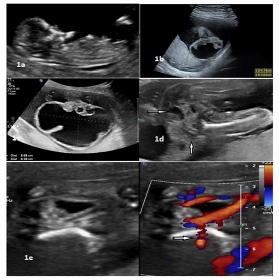
Case Report: We present a case of large fast growing (86x63mm) mostly cystic SCT in a 27-year-old woman with 23 weeks of gestational age and normal first trimester ultrasound exam. The fetus was evaluated by both magnetic resonance imaging (MRI) and ultrasonography.
Conclusion: Findings regarding tumor location, size, and content were similar for both sonography and MRI methods, though vascular pattern was detected with higher accuracy and more details by sonography. Meanwhile, MRI provided more appropriate information about tumor effects on surrounding tissue and conus location.


ارسال نظر