fetal lung maturation in fetuses with intrauterine growth restri
https://adir.tums.ac.ir//news/Comparison-of-fetal-lung-maturation-in-fetuses-with-intrauterine-growth-restriction-with-control-group,-using-lung-volume,-lung⁄liver-and-lung⁄muscle-signal-intensity-and-apparent-diffusion-coefficient-ratios-on-different-magnetic-resonance-imaging-sequences-
Comparison of ction with control group, using lung volume, lung/liver and lung/muscle signal intensity and apparent diffusion coefficient ratios on different magnetic resonance imaging sequences Purpose To compare lung volume, lung apparent diffusion coefficient (ADC) and signal intensity ratio (SIR) on different magnetic resonance imaging (MRI) sequences between intrauterine growth restriction (IUGR) fetuses and the control group. Materials and methods 49 IUGR and 58 non-IUGR fetuses were image

percutaneous transhepatic esophageal variceal
https://adir.tums.ac.ir//news/Multi-organ-infarction-following-percutaneous-transhepatic-esophageal-variceal-obliteration-with-glue-injection:-a-case-report
Multi organ infarction following obliteration with glue injection a case report Percutaneous transhepatic variceal obliteration (PTVO) is currently one of the best treatment options for controlling acute recurrent bleeding in cirrhotic patients. Nevertheless, this procedure is associated with major and minor complications such as fever, pain, fatal intraperitoneal hemorrhage, and rarely, embolization of embolic agents to the systemic circulation. Only one study has reported systemic emboli follo

Multi-parametric MRI for Parotid Tumors
https://adir.tums.ac.ir//news/Radiomic-Analysis-of-Multi-parametric-MR-Images-(MRI)-for-Classification-of-Parotid-Tumors-Running-Title:-Multi-parametric-MRI-for-Parotid-Tumors
Radiomic Analysis of Multi parametric MR Images (MRI) for Classification of Parotid Tumors Running Title Multi parametric MRI for Parotid Tumors Objective To determine the border of glial tumors by diffusion weighted imaging (DWI), apparent diffusion co-efficient (ADC), magnetic resonance spectroscopy (MRS) and perfusion brain MRI. Patients and methods Ten patients with brain gliomas were enrolled [mean age 35.3 ± 13.2, range 20-62]. Conventional MRI was performed for all patients. Beside
Perfusion Computed Tomography Scan Imaging in Differentiation of Benign from Malignant Parotid Lesions
https://adir.tums.ac.ir//Research-Article/Perfusion-Computed-Tomography-Scan-Imaging-in-Differentiation-of-Benign-from-Malignant-Parotid-Lesions
Introduction The most common site of salivary gland tumors is the parotid gland. Computed tomography (CT), magnetic resonance imaging (MRI), and sonography are imaging modalities to differentiate benign from malignant parotid tumors. Objective The aim of this study is the evaluation of the diagnostic value of perfusion CT for differentiating histological categorization of benign and malignant parotid tumors. Methods A total of 29 patients with parotid neoplasms were enrolled in

DWI in pelvic lymph nodes in endometrial cancer
https://adir.tums.ac.ir//news/Diagnostic-value-of-diffusion–weighted-magnetic-resonance-imaging-in-discriminating-between-metastatic-and-non–metastatic-pelvic-lymph-nodes-in-endometrial-cancer/270242
Diagnostic value of diffusion weighted magnetic resonance imaging in discriminating between metastatic and non metastatic pelvic lymph nodes in endometrial cancer Background Researchers have recently focused on assessing the accuracy of diffusion-weighted magnetic resonance imaging (DW-MRI) in predicting pelvic lymph node metastases in gynecological malignancies. Purpose To evaluate the diagnostic value of DW-MRI in discriminating between metastatic and non-metastatic pelvic lymph nodes in endom
.png)
Diagnostic Algorithm using Multi-parametric MRI Myometrial Tumo
https://adir.tums.ac.ir//article/A-Diagnostic-Algorithm-using-Multi-parametric-MRI-to-Differentiate-Benign-from-Malignant-Myometrial-Tumors:-Machine-Learning-Method
A Diagnostic Algorithm using Multi parametric MRI to Differentiate Benign from Malignant Myometrial Tumors Machine Learning Method This study aimed to develop a diagnostic algorithm for preoperative differentiating uterine sarcoma from leiomyoma through a supervised machine-learning method using multi-parametric MRI. A total of 65 participants with 105 myometrial tumors were included 84 benign and 21 malignant lesions (belonged to 51 and 14 patients, respectively; based on their postoperative ti

efficacy of (DWI) in of benign and malignant cervical lymphad
https://adir.tums.ac.ir//article/Diagnostic-efficacy-of-diffusion-weighted-magnetic-resonance-imaging-(DWI)-in-the-differentiation-of-benign-and-malignant-cervical-lymphadenopathies
Diagnostic efficacy of diffusion weighted magnetic resonance imaging (DWI) in the differentiation of benign and malignant cervical lymphadenopathies Objective To evaluate the diagnostic value of DWI in assessment of metastatic neck lymph node in a sample of Iranian patients with Head and Neck cancer. Methods 25 patients with 80 neck lymph nodes were analyzed using 1.5 T MRI. DWI was performed with b values of 0 and 1,000 s/mm2. Short axis diameter and ADC values (min, max and mean) were calculat
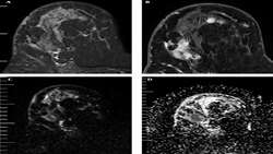
peritumoral edema with pathologic biomarkers in patients with br
https://adir.tums.ac.ir//Research-Article/Correlation-of-apparent-diffusion-coefficient-values-and-peritumoral-edema-with-pathologic-biomarkers-in-patients-with-breast-cancer
Correlation of apparent diffusion coefficient values and east cancer Purpose To investigate the relationship between breast cancer imaging features on magnetic resonance imaging (MRI) and histopathological characteristics. Methods and materials We prospectively enrolled 46 patients who underwent 1.5-T MRI with 68 breast malignant lesions from 2017 until 2019. Peritumoral edema was determined based on visual assessment on T2 weighted imaging. Lesions were categorized into two groups A with edema
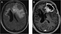
multimodal MR for differentiating infiltrative versus reactive
https://adir.tums.ac.ir//news/Evaluation-of-multimodal-MR-imaging-for-differentiating-infiltrative-versus-reactive-edema-in-brain-gliomas
Evaluation of multimodal MR imaging for differentiating infiltrative versus reactive edema in brain gliomas Objective To determine the border of glial tumors by diffusion weighted imaging (DWI), apparent diffusion co-efficient (ADC), magnetic resonance spectroscopy (MRS) and perfusion brain MRI. Patients and methods Ten patients with brain gliomas were enrolled [mean age 35.3 ± 13.2, range 20-62]. Conventional MRI was performed for all patients. Besides, tumor mapping based on Choline (Ch
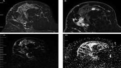
Peri-Tumoral Edema Determined in T2 Weighted Imaging
https://adir.tums.ac.ir//news/Correlation-of-Peri-Tumoral-Edema-Determined-in-T2-Weighted-Imaging-with-Apparent-Diffusion-Coefficient-of-Peritumoral-Area-in-Patients-with-Breast-Carcinoma
Correlation of Peri Tumoral Edema Determined in T2 Weighted Imaging with Apparent Diffusion Coefficient of Peritumoral Area in Patients with Breast Carcinoma Background Breast cancer may result in remodeling of adjacent normal appearing breast tissues. Magnetic resonance imaging (MRI) is increasingly used in the diagnosis and follow-up of breast cancer by means of diffusion weighted imaging, which is based on thermal motion of water molecules in the extracellular fluid. Objectives We investiga