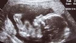
مقایسه حجم، سیگنال و ADC ریه درmri جنین های نرمال و جنین IUGR
https://adir.tums.ac.ir//projects/مقایسه-حجم،-سیگنال-و-ADC-ریه-در-ام-آر-آی-جنین-های-نرمال-و-جنین-های-مبتلا-به-IUGR-در-رنج-سنی-مشابه،-مراجعه-کننده-به-بیمارستان-یاس-و-امام-خمینی-در-طی-سال-های-99⁄1398
مقایسه حجم، سیگنال و ADC ریه در ام آر آی جنین های نرمال و جنین های مبتلا به IUGR در رنج سنی مشابه، مراجعه کننده به بیمارستان یاس و امام خمینی در طی سال های 99/1398 کد خبر 264775 کلمات کلیدی iugr_adc_mri مدیر سایت *

دقت تشخیصی (DWI) و (ADC) جهت تعیین بدخیمی توده های آدنکسال
https://adir.tums.ac.ir//projects/بررسی-دقت-تشخیصی-Diffusion–weighted-imaging(DWI)-و-apparent-diffusion-coefficient-(ADC)-value--جهت-تعیین-بدخیمی-توده-های-آدنکسال-مراجعه-کننده-به-بخش-جراحی-زنان-بیمارستان-ولیعصر-(عج)-در-سال-1393
بررسی دقت تشخیصی Diffusion-weighted imaging(DWI) و apparent diffusion coefficient (ADC) value جهت تعیین بدخیمی توده های آدنکسال مراجعه کننده به بخش جراحی زنان بیمارستان ولیعصر (عج) در سال 1393 کد خبر 263852 کلمات کلیدی dwiadc مدیر سایت *

ADC-derived spatial features can classify adnexal lesions
https://adir.tums.ac.ir//article/ADC-derived-spatial-features-can-accurately-classify-adnexal-lesions
ADC derived spatial features can accurately classify adnexal lesions Background The role of quantitative apparent diffusion coefficient (ADC) maps in differentiating adnexal masses is unresolved Purpose/hypothesis To propose an objective diagnostic method devised based on spatial features for predicting benignity/malignancy of adnexal masses in ADC maps Study type Prospective Population In all, 70 women with sonographically indeterminate and histopathologically confirmed adnexal masses (38 benig

Weighted Imaging and Proton MRS Findings in Osteosarcoma
https://adir.tums.ac.ir//news/Weighted-Imaging-and-Proton-Magnetic-Resonance-Spectroscopy-Findings-in-Osteosarcoma-Versus-Normal-Muscle
Weighted Imaging and Proton Magnetic Resonance Spectroscopy Findings in Osteosarcoma Versus Normal Muscle Objectives The goal of our study was to assess diffusion-weighted (DW) imaging and proton magnetic resonance (MR) spectroscopy findings in osteosarcoma versus normal muscle at 3 Tesla (3 T) MR system. Patients and Methods Nineteen patients highly suspicious for osteosarcoma and 12 normal young healthy adults were enrolled in this study. Two patients were excluded from the study due to incomp
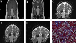
Tumor in Pituitary Macroadenomas & Preoperative Diffusion (ADC)
https://adir.tums.ac.ir//news/-Is-There-Any-Congruity-Between-Tumor-Consistency-in-Pituitary-Macroadenomas-and-Preoperative-Diffusion-Weighted-Imaging-and-Apparent-Diffusion-Coefficient-(ADC)-Map؟
Is There Any Congruity Between Tumor Consistency in Pituitary Macroadenomas and Preoperative Diffusion Weighted Imaging and Apparent Diffusion Coefficient (ADC) Map? Background Pituitary adenomas are the most common space-occupying lesions in the sellar region. Pituitary adenomas are usually soft, but 5 to 13% of macroadenomas are fibrous. The resection of firm tumors by curettage or suction is challenging. Therefore, preoperative knowledge of the pituitary adenomas consistency could help neuros

Differentiation of benign from malignant adnexal masses
https://adir.tums.ac.ir//article/Differentiation-of-benign-from-malignant-adnexal-masses-by-functional-3-tesla-MRI-techniques:-diffusion-weighted-imaging-and-time-intensity-curves-of-dynamic-contrast-enhanced-MRI
by functional 3 tesla MRI techniques diffusion weighted imaging and time intensity curves of dynamic contrast enhanced MRI The aim of this study was to evaluate and compare the accuracy of diffusion-weighted imaging (DWI), apparent diffusion coefficient (ADC) value, and time-intensity curve (TIC) type analysis derived from dynamic contrast-enhanced MR imaging (DCE-MRI) in differentiating benign from malignant adnexal masses Background The aim of this study was to evaluate and compare the accurac
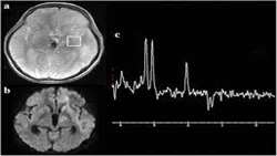
حساسیت و اختصاصیت ترکیب دو روش (MRS) و DWI در درجه بندی تومورهای
https://adir.tums.ac.ir//projects/-بررسی-حساسیت-و-اختصاصیت-ترکیب-دو-روش-(MRS)-و-DWI-در-درجه-بندی-تومورهای-گلیال
بررسی گلیال در مطالعه حاضر کلیه بیماران مراجعه کننده به درمانگاه جراحی اعصاب با تشخیص گلیوم مغزی بر اساس نظر نورورادیولوژیست مجرب با توجه به کلیشه MRI که کاندید جراحی هستند تحت بررسی قرار گرفتند. بیماران با تومورهای گلیال اینفراتنتوریال که امکان مطالعه دقیق انها بکمک MRS و DWI کمتر فراهم است , انهایی که قبل از انجام مطالعه تحت مداخله درمانی ماژور قرار گرفته اند و از طرفی بیمارانی که با فاصله بیش از 1 ماه از زمان انجام مطالعه تحت جراحی قرار گرفته اند , از مطالعه خارج شدند. تومورهای مورد مطالعه بر