DWI and IVIM parameters in differentiating intrahepatic cholangi
https://adir.tums.ac.ir//article/Diagnostic-value-of-quantitative-DWI-and-IVIM-parameters-in-differentiating-intrahepatic-cholangiocarcinoma-and-hepatocellular-carcinoma:-a-systematic-review-and-meta–analysis
Diagnostic value of quantitative ocarcinoma and hepatocellular carcinoma a systematic review and meta-analysis Purpose This study evaluates the diagnostic performance of quantitative diffusion-weighted imaging (DWI), including both conventional and intravoxel incoherent motion (IVIM)-derived parameters, in differentiating hepatocellular carcinoma (HCC) from intrahepatic cholangiocarcinoma (ICC). Method A systematic review and meta-analysis were conducted following a pre-registered protocol ( htt
O-RADS MRI evaluation for characterizing ovarian masses
https://adir.tums.ac.ir//news/The-added-value-of-apparent-diffusion-coefficient-assessments-in-O–RADS-MRI-evaluation-for-characterizing-ovarian-masses-with-solid-components
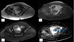
The added value of apparent diffusion coefficient assessments in with solid components Abstract Background Integrating diffusion-weighted imaging (DWI) and apparent diffusion coefficient (ADC) measurements with existing MR imaging protocols improves the differentiation between benign and malignant adnexal lesions. We aimed to assess the additional value of quantitative ADC in diagnosing adnexal masses classified by the O-RADS-MRI score and evaluate the impact on diagnostic performance. Methods T

Apparent Diffusion Coefficient MRI in Hepatocellular Adenoma
https://adir.tums.ac.ir//article/Comparing-Texture-Analysis-of-Apparent-Diffusion-Coefficient-MRI-in-Hepatocellular-Adenoma-and-Hepatocellular-Carcinoma
Comparing Texture Analysis of and Hepatocellular Carcinoma Aim This study aimed to assess the effectiveness of using MRI-apparent diffusion coefficient (ADC) map-driven radiomics to differentiate between hepatocellular adenoma (HCA) and hepatocellular carcinoma (HCC) features. Materials and methods The study involved 55 patients with liver tumors (20 with HCA and 35 with HCC), featuring 106 lesions equally distributed between hepatic carcinoma and hepatic adenoma who underwent texture analysis o

DTI for Diagnosing and Assessing Cervical Myelopathy
https://adir.tums.ac.ir//article/Diffusion-Tensor-Imaging-in-Diagnosing-and-Evaluating-Degenerative-Cervical-Myelopathy-A-Systematic-Review-and-Meta-Analysis
Diffusion Tensor Imaging in Diagnosing and Evaluating Degenerative Cervical Myelopathy A Systematic Review and Meta Analysis Objective Degenerative cervical myelopathy (DCM) is a common spinal cord disorder necessitating surgery. We aim to explore how effectively diffusion tensor imaging (DTI) can distinguish DCM from healthy individuals and assess the relationship between DTI metrics and symptom severity. Methods We included studies with adult DCM patients who had not undergone decompressive su

مقایسه ADC در SCC، لنفوم و بافت نرمال
https://adir.tums.ac.ir//news/بررسی-و-مقایسه-کمیتهای-گوناگون-ADC-بین-بافتهای-کارسینوم-سلولهای-سنگفرشی(SCC)،-لنفوم-و-بافت-لنفوئیدی-نرمال-در-تصاویر-MRI-سر-و-گردن
بررسی و مقایسه کمیتهای گوناگون ADC بین بافتهای کارسینوم سلولهای سنگفرشی(SCC)، لنفوم و بافت لنفوئیدی نرمال در تصاویر MRI سر و گردن کد خبر 293311 کلمات کلیدی adc_سلولهای_سنگفرشیmri_ مدیر سیستم 0 * *
ارزش تشخیصی DWIو ADC و perfusion MRI و MRS در افتراق ادم ریکتیو
https://adir.tums.ac.ir//projects/بررسی-ارزش-تشخیصی-DWIو-ADC-و-perfusion-MRI-و-MRS-در-افتراق-ادم-ریکتیو-از-ادم-اینفیلتراتیو-در-بیماران-مبتلا-به-گلیومای-مغزی
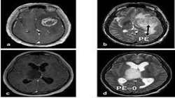
بررسی از ادم اینفیلتراتیو در بیماران مبتلا به گلیومای مغزی کد خبر 293239 کلمات کلیدی تومور_گلیوماادمadc_dwimrs_ مدیر سایت 0 * *
ارزش تشخیصی پارامترهایDwi, ADC value,T2 signal ,Perfusion MR
https://adir.tums.ac.ir//done-projects/بررسی-ارزش-تشخیصی-پارامترهایDiffusion-weighted-MRI,-ADC-value,T2-signal-,Perfusion-weighted-MR-حهت-افتراق-سارکوم-رحمی-از-میوم
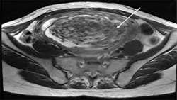
بررسی ارزش تشخیصی پارامترهایDiffusion weighted MRI, ADC value,T2 signal ,Perfusion weighted MR حهت افتراق سارکوم رحمی از میوم کد خبر 293237 کلمات کلیدی سارکوم_رحمیمیوم مدیر سیستم 0 * *
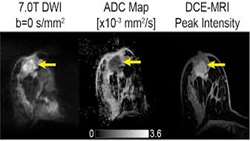
ADC در بررسیDWI ضایعات خوش خیم و بدخیم پستان
https://adir.tums.ac.ir//news/مقایسه-عدد-ADC-در-بررسی-Diffusion–weighted-imagining-(DWI)-ضایعات-خوش-خیم-و-بدخیم-پستان-و-تعین-حساسیت-و-ویژگی-DWI-در-این-خصوص·
مقایسه عدد ADC در بررسی Diffusion-weighted imagining (DWI) ضایعات خوش خیم و بدخیم پستان و تعین حساسیت و ویژگی DWI در این خصوص. کد خبر 293232 کلمات کلیدی dwiadc_ضایعات_پستان مدیر سیستم 0 * *

Benign hepatic schwannoma in chronic hepatitis B infection
https://adir.tums.ac.ir//case-report/Benign-hepatic-schwannoma-in-a-patient-with-chronic-hepatitis-B-infection:-A-case-report
Benign hepatic schwannoma in a patient with chronic hepatitis B infection A case report A primary benign hepatic schwannoma is an extremely rare disease with a good prognosis. A 55-year-old man with chronic hepatitis B was referred to our hospital because of jaundice, weight loss, and a hepatic lesion found during an ultrasound examination. Magnetic resonance image revealed a 55 × 120 mm solid mass lesion in the segment V and VIII of the liver. The mass extended directly to the segmental b

Liver hydatidosis disease with portal vein invasion
https://adir.tums.ac.ir//case-report/Liver-hydatidosis-disease-with-portal-vein-invasion:-Report-of-a-rare-case-and-review-of-literature
Report of a rare case and review of literature Hydatid disease is a zoonosis caused by Echinococcus granulosus. Humans are aberrant intermediate hosts and following the infection, the parasite may infest any organ of the body, with the liver and lungs being the most involved organs. Portal vein involvement by hydatid cyst disease is extremely rare with only seven cases published to our knowledge. We present a 62-year-old Persian male with an incidental liver cyst. His laboratory tests were in no