Pelvic MRI for Depth of Myometrial Invasion and Cervical Involve
https://adir.tums.ac.ir//article/Diagnostic-Value-of-Pelvic-MRI-for-Assessment-of-the-Depth-of-Myometrial-Invasion-and-Cervical-Involvement-in-Endometrial-Cancer:-Comparison-of-New-Versus-Old-FIGO-Staging-
Diagnostic Value of Pelvic MRI for Assessment of the Depth of Myometrial Invasion and Cervical Involvement in Endometrial Cancer Comparison of New Versus Old FIGO Staging Using 2009 FIGO classification increases the accuracy of pelvic MR imaging for preoperative staging of patients with early stages of endometrial cancer BackgroundEndometrial carcinoma is a highly prevalent gynecologic malignancy. The International Federation of Gynecology and Obstetrics (FIGO) staging system underwent significa

Chemoembolization on Lesion Reduction in Liver Metastases
https://adir.tums.ac.ir//article/Efficacy-of-Transarterial-Chemoembolization-on-Lesion-Reduction-in-Colorectal-Liver-Metastases
Efficacy of Transarterial Chemoembolization on Lesion Reduction in Colorectal Liver Metastases The aim of present study was to evaluate the efficacy of chemoembolization for inoperable metastatic liver lesions from CRC Following failure of systemic chemotherapy, transarterial chemoembolization (TACE) is an available method to control unresectable liver metastases from colorectal carcinoma (CRC). The aim of present study was to evaluate the efficacy of chemoembolization for inoperable metastatic
.jpg)
Imaging-Guided Interventions in Management of Hepatocellular Car
https://adir.tums.ac.ir//article/Imaging-and-Imaging-Guided-Interventions-in-the-Diagnosis-and-Management-of-Hepatocellular-Carcinoma-(HCC)-Review-of-Evidence
Imaging and Imaging Guided Interventions in the Diagnosis and Management of Hepatocellular Carcinoma (HCC) Review of Evidence imaging (mainly CT and MR imaging) has a crucial role in the diagnosis and assessment of tumor response in patients with HCC and interventional therapies play a crucial role in the management of those who are not candidates for surgical therapies. The imaging of hepatocellular carcinoma (HCC) is challenging and plays a crucial role in the diagnosis and staging of the dise
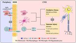
Lipoic acid and multiple sclerosis
https://adir.tums.ac.ir//article/Lipoic-acid-and-multiple-sclerosis:-A-randomized-controlled-conical-trial
A randomized controlled conical trial Objective To determine whether lipoic acid (LA), an endogenously produced antioxidant, slowed the whole-brain atrophy rate and was safe in secondary progressive MS (SPMS). Methods Patients with SPMS aged 40-70 years enrolled in a single center, 2-year, double-blind, randomized trial of daily oral 1,200 mg LA vs placebo. Primary outcome was change in annualized percent change brain volume (PCBV). Secondary outcomes were changes in rates of atrophy of segmente
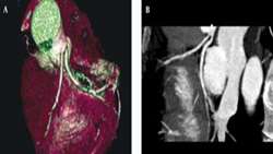
Congenital Coronary Artery Anomalies using Coronary CTAngiograph
https://adir.tums.ac.ir//Research-Article/Prevalence-of-Congenital-Coronary-Artery-Anomalies-and-Variants-in-2697-Consecutive-Patients-Using-64-Detector-Row-Coronary-CTAngiography
Prevalence of Congenital Coronary Artery Anomalies and Variants in 2697 Consecutive Patients Using 64 Detector Row Coronary CTAngiography Based on the fact that coronary CT-angiography using MDCT can display different coronary anomalies, this study shows similar results to other reports on the subject Background Coronary artery anomalies are not common, but could be very serious. Objectives This study determines the frequency of coronary anomalies and normal variants by multi-detector-row comput
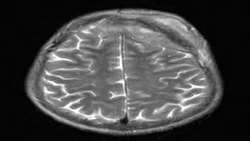
diffuse large B-cell non-Hodgkin lymphoma of the cranial vault
https://adir.tums.ac.ir//Research-Article/Primary-diffuse-large-B-cell-non-Hodgkin-lymphoma-of-the-cranial-vault
Primary diffuse large B cell non Hodgkin lymphoma of the cranial vault Primary non-Hodgkin’s lymphoma of the cranial vault is extremely rare Primary non-Hodgkin’s lymphoma of the cranial vault is extremely rare. This case report presents a 42-year-old man with a painless subcutaneous scalp mass which extended intracranially associated with recent mild headache. Initial computed tomography and magnetic resonance imaging revealed two lesions emanating from the skull. Biopsy revealed a diagno
carotid advanced atherosclerotic stenosis
https://adir.tums.ac.ir//Research-Article/Quantitative-T2*-magnetic-resonance-imaging-for-evaluation-of-iron-deposition-in-the-brain-of-β-thalassemia-patients
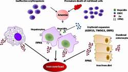
Quantitative T2* magnetic resonance imaging for evaluation of iron deposition in the brain of β thalassemia patients Serum ferritin and liver iron content may not be good indicators of brain iron deposition in patients with β thalassemia major. Background Iron overload is a common clinical problem in patients with β-thalassemia major. The purpose of this study was to assess the presence of excess iron in certain areas of the brain (thalamus, midbrain, adenohypophysis and basal ganglia) in p
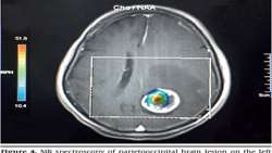
diagnostic accuracy of cystic brain tumors in 3 tesla MRI
https://adir.tums.ac.ir//article/The-effects-of-voxel-localization-and-time-of-echo-on-the-diagnostic-accuracy-of-cystic-brain-tumors-in-3-tesla-magnetic-resonance-spectroscopy
The effects of voxel localization and time of echo on the diagnostic accuracy of cystic brain tumors in 3 tesla magnetic resonance spectroscopy Selection of voxels with the guide of chemical shift imaging map yields to 100% diagnostic sensitivity. If not accessible, the use of the union of peripheral and central voxels enhances the sensitivity when compared to usage of peripheral or central voxels solely. Background Although magnetic resonance spectroscopy (MRS) has been shown as an effective di

The Prospective Profile of Endovascular Coiling of Cerebral Aneu
https://adir.tums.ac.ir//aneurym/The-Prospective-Profile-of-Endovascular-Coiling-of-Cerebral-Aneurysms:-The-Impact-of-Clinical-Presentation,-Procedural-Results,-Aneurysm-Rupture-and-Location
rysms The Impact of Clinical Presentation, Procedural Results, Aneurysm Rupture and Location Selection of voxels with the guide of chemical shift imaging map yields to 100% diagnostic sensitivity. If not accessible, the use of the union of peripheral and central voxels enhances the sensitivity when compared to usage of peripheral or central voxels solely. Background Although magnetic resonance spectroscopy (MRS) has been shown as an effective diagnostic tool in distinguishing inflammation from n
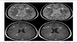
acute phase of relapsing remitting multiple sclerosis
https://adir.tums.ac.ir//Research-Article/The-relationship-between-enhanced-plaques-with-Gadovist-and-Magnevist-contrast-brain-magnetic-resonance-imaging-and-the-neurological-deficit-in-the-acute-phase-of-relapsing-remitting-multiple-sclerosis
The relationship between enhanced plaques with Gadovist and Magnevist contrast brain magnetic resonance imaging and the neurological deficit in the both Magnevist and Gadovist could be used as the contrast material to detect enhancing plaques in relapse phase of multiple sclerosis Background Magnetic resonance imaging (MRI) is the standard method for observing brain plaques and contrast material injection is necessary for demonstrating the active plaque.This study compared the rate of enhancemen