Coronary CTA and Dual-Energy CT in Ischemic Heart
https://adir.tums.ac.ir//news/Coronary-CT-Angiography-and-Dual-Energy-Computed-Tomography-in-Ischemic-Heart-Disease-Suspected-Patients
Coronary CT Angiography and Dual Energy Computed Tomography in Ischemic Heart Disease Suspected Patients Background Advanced computed tomography (CT) scanners enable concurrent assessment of coronary artery anatomy and myocardial perfusion. The purpose of this study was to assess dual-energy CT images in a group of patients suspected for ischemic heart disease and to evaluate agreement of cardiac computed tomography perfusion (CTP) images with CT angiography results in a single dual-energy compu
Diagnostic of MRI and Echocardiography in Constrictive Pericardi
https://adir.tums.ac.ir//article/Diagnostic-Efficacy-of-Magnetic-Resonance-Imaging-and-Echocardiography-in-Diagnosis-of-Constrictive-Pericarditis
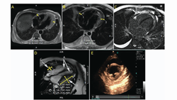
Diagnostic Efficacy of Magnetic Resonance Imaging and Echocardiography in Diagnosis of Constrictive Pericarditis Breast intraductal lesions present a diagnostic challenge due to the diverse spectrum of histologic changes. Vacuum-assisted biopsy (VAB) has evolved as a pivotal diagnostic and therapeutic modality. Yet, concerns about the underestimation of malignancy using VAB persist. This review examines the underestimation rates of Ultrasound-guided VAB (US-VAB) for intraductal lesions and evalu
contrast dose and diffusion coefficient measurement on vessel s
https://adir.tums.ac.ir//news/Evaluation-of-contrast-agent-dose-and-diffusion-coefficient-measurement-on-vessel-size-index-estimation
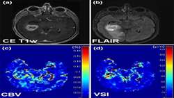
Evaluation of contrast agent dose and diffusion coefficient measurement on vessel size index estimation Objectives The goal of this study is to examine the effect of contrast agent (CA) dose and diffusion coefficient on the estimation of vessel size index (VSI). Materials and methods Three groups of four participants were enrolled in this study and two different experiments were performed. Different dose of CA, namely 0.1 mmol/kg and 0.05 mmol/kg were assessed in two groups of normal subjects. D

progressive multifocal leukoencephalopathy after liver transpla
https://adir.tums.ac.ir//news/A-41-year-old-female-with-progressive-multifocal-leukoencephalopathy-after-liver-transplant
A 41 year old female withnt Progressive multifocal leukoencephalopathy (PML) is a demyelinating disease caused by JC virus reactivation. Its occurrence is very rare after solid organ transplantation, especially liver transplantation. We report a patient who received liver transplantation due to liver failure resulting from autoimmune hepatitis and advanced PML presenting with aphasia. A 41-year-old female with a history of liver transplantation who received a usual immunosuppression regimen was
Design and Validation of Synchronous QCT Calibration Phantom
https://adir.tums.ac.ir//article/Design-and-Validation-of-Synchronous-QCT-Calibration-Phantom:-Practical-Methodology
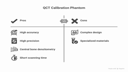
Practical Methodology Introduction Quantitative computed tomography (QCT) can supplement dual x-ray absorptiometry by enabling geometric and compartmental bone assessments. Whole-body spiral CT scanners are widely available and require a short scanning time of seconds, in contrast to peripheral QCT scanners, which require several minutes of scanning time. This study designed and evaluated the accuracy and precision of a homemade QCT calibration phantom using a whole-body spiral CT scanner. Mater

low back pain with unilateral radiculopathy caused by disc hern
https://adir.tums.ac.ir//news/Neuromuscular-morphometric-characteristics-in-low-back-pain-with-unilateral-radiculopathy-caused-by-disc-herniation:-An-ultrasound-imaging-evaluation
Neuromuscular morphometric characteristics iniation An ultrasound imaging evaluation Background Little is known about the neuromuscular morphometric characteristics in patients with sciatica. Objective To evaluate the possible changes of nerve and muscle structures in patients with low back pain with unilateral radiculopathy due to lumbar disc herniation by ultrasound imaging. Design A case-control observational study. Methods Forty individuals were divided into case (n = 20; low back pain with

3T DCEMRI Parameters as Predictors of Malignancy in Adnexal
https://adir.tums.ac.ir//article/Comparison-of-Qualitative-(Time-Intensity-Curve-Analysis),-Semi-Quantitative,-and-Quantitative-Multi-Phase-3T-DCEMRI-Parameters-as-Predictors-of-Malignancy-in-Adnexal
Comparison of Qualitative (Time Intensity Curve Analysis), Semi Quantitative, and Quantitative Multi Phase Objective The present study aimed to compare the qualitative (time intensity curve analysis), the semi-quantitative and the quantitative multiphase 3T dynamic contrast-enhanced (DCE) MRI parameters as predictors of malignancy in adnexal masses. Materials and Methods In this prospective study, women with an adnexal mass who were scheduled for surgical resection or were followed for more than

Sonoelastography for skin evaluation in sclerodermic patients
https://adir.tums.ac.ir//article/Sonoelastography-for-skin-evaluation-in-sclerodermic-patients
Background The objective of the study is to evaluate elastography ultrasound findings in patients with scleroderma (SS) and to clarify the effectiveness of elastosonography to differentiate scleroderma lesions from any skin lesion considering tissue elasticity. Methods Thirty-six SS patients definite diagnosis of systemic sclerosis according to American College of Rheumatology criteria and 36 healthy subjects were enrolled. Volar aspect of the middle forearm and arm in addition to the dorsal asp

Pregnant Trauma Patient with Asymptomatic Fat Embolism
https://adir.tums.ac.ir//case-report/A-21-year-old-Pregnant-Trauma-Patient-with-Asymptomatic-Fat-Embolism-a-Case-Report
A 21 year old a Case Report Introduction Fat embolism syndrome (FES) is most often associated with orthopedic trauma that typically presents 24–72 hours after the trauma with a classic triad. Only few cases of fat embolism due to lower extremity venous system had been reported. Case presentation The current case report presents a pregnant woman who was referred to our emergency department with bilateral femoral open fracture. After detecting fetal demise by abdominopelvic ultrasound, an ar

Fluoroscopic analysis of anterior tibial translation
https://adir.tums.ac.ir//article/Fluoroscopic-analysis-of-anterior-tibial-translation-during-eccentric-and-concentric-phase-of-knee-rehabilitation-exercises-in-men-with-anterior-cruciate-ligament-injury
during eccentric and concentric phase of knee rehabilitation exercises in men with anterior cruciate ligament injury Background The amount of anterior tibial translation during rehabilitation exercises is a key factor in organizing exercise regimen after anterior cruciate ligament injury. Excessive anterior tibial translation could increase the magnitude of tension imposed on injured and reconstructed anterior cruciate ligament knees. Forward lunge and open-kinetic knee extension exercises are c