Ultrasound and shear-wave elastography for treatment of morphe
https://adir.tums.ac.ir//Research-Article/Ultrasound-and-shear–wave-elastography-as-a-tool-for-evaluating-response-to-treatment-of-morphea:-a-preliminary-report-of-a-prospective-study
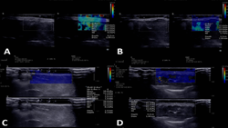
Ultrasound and shear-wave elastography as a tool for evaluating response to treatment of morphea a preliminary report of a prospective study Morphea is an autoimmune connective tissue disorder with no established objective biomarkers for treatment monitoring. Due to the subjectivity of current clinical scoring systems, this study investigates the efficacy of grayscale ultrasound, color Doppler, and shear-wave elastography (SWE) in assessing treatment response and correlating with clinical scores
Elastography for distinguishing lymphoma from lymphadenopathy
https://adir.tums.ac.ir//news/Elastography-for-distinguishing-lymphoma-from-benign-and-metastatic-lymphadenopathy:-a-systematic-review-and-Bayesian-meta–analysis/331985
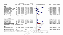
Elastography for distinguishing lymphoma from benign and metastatic lymphadenopathy a systematic review and Bayesian meta-analysis Background Differentiating lymphoma from benign and metastatic lymphadenopathies using conventional ultrasound remains clinically challenging. Despite the utility of ultrasound elastography in assessing tissue stiffness, the intermediate stiffness profile of lymphoma frequently leads to misclassification. To address this diagnostic gap, we conducted a comprehensive s
Transperineal strain elastography for evaluating periurethral bi
https://adir.tums.ac.ir//news/Transperineal-strain-elastography-for-evaluating-periurethral-biomechanics-in-women-with-stress-urinary-incontinence:-a-diagnostic-accuracy-study/331928
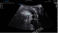
omechanics in women with stress urinary incontinence a diagnostic accuracy study Background Stress urinary incontinence (SUI) commonly reflects loss of urethral support and altered periurethral biomechanics. Strain elastography (SE) can quantify tissue deformation; however, its clinical diagnostic value for SUI is uncertain. Objective To evaluate periurethral strain elastography in women with and without SUI and to compare its diagnostic performance with established kinematic ultrasound paramete

Doppler US and US Elastography is Superior to Doppler US or US
https://adir.tums.ac.ir//Research-Article/Combination-of-Doppler-US-and-US-Elastography-is-Superior-to-Doppler-US-or-US-Elastography-Alone-in-Detecting-Delayed-Kidney-Graft-Rejection
Combination of Elastography Alone in Detecting Delayed Kidney Graft Rejection Objectives To compare the performance of Doppler ultrasound (US) and US elastography with their combination in detecting delayed graft rejection. Methods A prospective cross-sectional study of 60 consecutive adult kidney transplant recipients was done. Patients with creatinine > 1.5 mg/dL and a minimum interval of 3 months from renal transplant surgery were recruited. All patients underwent both Doppler US and US el

ultrasound elastography for uterine cervical neoplasms
https://adir.tums.ac.ir//article/Diagnostic-and-clinical-utility-of-ultrasound-elastography-for-uterine-cervical-neoplasm
Diagnostic and clinical utility of ultrasound elastography for uterine cervical neoplasm Purpose Cervical cancer remains a major burden, especially in low-resource settings, due to limited screening. Traditional diagnostic tools, like colposcopy, are operator-dependent and often lack accuracy. Ultrasound elastography, including strain elastography (SE) and shear wave elastography (SWE), is a cost-effective, non-invasive tool for assessing tissue stiffness, aiding in distinguishing benign from ma

Sonographic skin features and shear wave elastography
https://adir.tums.ac.ir//news/Sonographic-skin-features-and-shear-wave-elastography-in-distinguishing-active-from-inactive-morphea-lesions:-A-case–control-study/292774
in distinguishing active from inactive morphea lesions A case control study Morphea (localized scleroderma) is a connective tissue disorder characterized by dermal stiffness, thickening, erythema, and pigmentation abnormalities, often varying with the degree of inflammation and fibrosis.1 Precise lesion activity is crucial for tailoring optimal treatment plans. Traditional diagnostic methods like skin biopsy are invasive and may not effectively capture the dynamic progression of the disease.2 No

Application of Sonoelastography in Differential Diagnosis Thyroi
https://adir.tums.ac.ir//news/Application-of-Sonoelastography-in-Differential-Diagnosis-of-Benign-and-Malignant-Thyroid-Nodules
Application of Sonoelastography in Differential Diagnosis of Benign and Malignant Thyroid Nodules Sonoelastography could help to differentiate benign and malignant thyroid nodules. As our sample size was limited, larger studies are recommended Background Sonoelastography is a new ultrasound method which could be helpful to determine which thyroid nodule is malignant. We designed this study to evaluate the accuracy of sonoelastography in differentiating of benign and malignant thyroid nodules in

ultrasound elastography for determining carpal tunnel syndrome
https://adir.tums.ac.ir//news/Application-of-ultrasound-elastography-for-determining-carpal-tunnel-syndrome-severity
Application of severity To evaluate grey-scale and elastography ultrasound imaging findings in patients with CTS compared to nerve conductive studies. Objective To evaluate grey-scale and elastography ultrasound imaging findings in patients with CTS compared to nerve conductive studies. Method Sixty median nerves of 31 cases with confirmed CTS and 44 median nerves in 22 controls (healthy volunteers) who had no clinical evidence of CTS were evaluated. An expert radiologist performed all US evalua

Sonoelastography in Addition to Ultrasound in Investigating Brea
https://adir.tums.ac.ir//news/Diagnostic-Performance-of-Sonoelastography-in-Addition-to-Ultrasound-in-Investigating-Breast-Lesions:-Can-Concomitant-Use-of-These-Techniques-Lead-to-Improvement-of-Differentiation
Diagnostic Performance of st Lesions Can Concomitant Use of These Techniques Lead to Improvement of Differentiation simultaneous evaluation of suspicious breast lesions with both US and SE can have high sensitivity and specificity and prevent the unnecessary invasive interventions. Background Sonoelastography (SE) is introduced as a complementary technique for ultrasoungraohy (US) to evaluate breast lesions. This method is based on tissue strain in response to compression and decompression. The