contrast-enhanced mammography in evaluating breast cancer respon
https://adir.tums.ac.ir//Research-Article/Diagnostic-accuracy-of-contrast–enhanced-mammography-in-evaluating-breast-cancer-response-to-neoadjuvant-therapy:-a-systematic-review-and-meta–analysis
Diagnostic accuracy of se to neoadjuvant therapy a systematic review and meta-analysis Purpose Neoadjuvant therapy (NAT) in breast cancer enables tumor downstaging and response evaluation. Although magnetic resonance imaging (MRI) is the gold standard, its limitations have prompted growing interest in alternatives such as contrast-enhanced mammography (CEM). Methods This review was conducted according to PRISMA guidelines. We evaluated CEM accuracy in predicting pathological complete response af

Breast arterial calcifications on mammography and risk of stroke
https://adir.tums.ac.ir//Research-Article/Breast-arterial-calcifications-on-mammography-and-risk-of-stroke:-a-systematic-review-and-meta–analysis
a systematic review and meta-analysis Background Breast arterial calcifications (BAC) are commonly observed as an incidental finding on screening mammography and have been linked to cardiovascular disease. Whether BAC is independently associated with stroke risk remains uncertain. Methods We conducted a systematic review and meta-analysis in accordance with PRISMA 2020 guidelines. PubMed, Embase, and Scopus were searched from inception to May 2024 for cohort studies evaluating the association be
.png)
Diagnostic Probe vs. Mammography Accuracy in Cancer Surgery
https://adir.tums.ac.ir//news/Evaluation-of-the-Concordance-of-Cancer-Diagnostic-Probe-Findings-During-Surgery-and-Suspected-Distortion-in-Pre-surgical-Mammography-Based-on-Pathology
Evaluation of the Concordance of Cancer Diagnostic Probe Findings During Surgery and Suspected Distortion in Pre surgical Mammography Based on Pathology Cancer diagnostic probe stations (CDP) are expensive, and they are better used in patients with suspicious findings in initial mammography. For the first time, this study investigated the compatibility of suspicious findings observed in mammograms of patients before surgery with CDP findings based on pathology findings as the gold standard. This

Breast Cancer Screening Based on Mammography
https://adir.tums.ac.ir//news/Diagnostic-Accuracy-of-Opportunistic-Breast-Cancer-Screening-Based-on-Mammography-in-Iran
Diagnostic Accuracy of Opportunistic in Iran Background Although breast cancer is the most prevalent type of cancer among Iranian women, its screening is opportunistic in Iran. The available guidelines for breast cancer screening are based on mammography. A screening modality should have adequate diagnostic accuracy and be widely available at reasonable cost. Although mammography is highly accessible in Iran, its accuracy has not been investigated. Objectives This study aimed to evaluate the acc
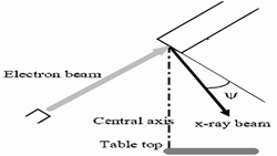
Calculation of the contrast of the calcification in digital mamm
https://adir.tums.ac.ir//news/Calculation-of-the-contrast-of-the-calcification-in-digital-mammography-system:-Gate-validation
ography system Gate validation Purpose Validation of the Gate tool in digital mammography image simulation from the viewpoint of image quality (contrast of calcifications). Materials and methods The polymethyl methacrylate (PMMA) phantom containing aluminum foils in different thicknesses is used for measuring the contrast of calcifications in a real system. In this research, the phantom and mammography system have been simulated by the Gate tool with the maximum possible details. The contrast of

Ultra-Fast Image Reconstruction of Tomosynthesis Mammography Usi
https://adir.tums.ac.ir//news/Ultra-Fast-Image-Reconstruction-of-Tomosynthesis-Mammography-Using-GPU
Ultra Fast Image Reconstruction of Tomosynthesis Mammography Using GPU This research has been designed to study ultra-fast image reconstruction technique for Tomosynthesis Mammography systems using Graphics Processing Unit (GPU) Digital Breast Tomosynthesis (DBT) is a technology that creates three dimensional (3D) images of breast tissue. Tomosynthesis mammography detects lesions that are not detectable with other imaging systems. If image reconstruction time is in the order of seconds, we can u