


Diagnostic utility of quantitative susceptibility mapping in Parkinson’s disease and atypical parkinsonism: A systematic review and meta-analysis
Background: Quantitative susceptibility mapping (QSM) is an advanced MRI technique that measures tissue magnetic susceptibility, serving as a non-invasive biomarker for iron content. It has shown increasing promise for differentiating idiopathic Parkinson's disease (PD) from atypical Parkinsonian syndrome (APS), including multiple system atrophy (MSA), progressive supranuclear palsy (PSP). This systematic review and meta-analysis evaluated the diagnostic utility of QSM in Parkinsonian syndromes.
Purpose: We systematically searched PubMed, Scopus, Web of Science, and Embase through November 2025 for observational studies reporting QSM metrics in patients with Parkinsonian disorders and healthy controls. Standardized mean differences (SMDs) with 95% confidence intervals (CIs) were pooled for regional susceptibility values using random-effects models. When available, sensitivity, specificity, and area under the receiver operating characteristic curve (AUC) were synthesized using bivariate random-effects models. Heterogeneity was quantified with I² and explored with subgroup analyses and meta-regression. Publication bias was assessed with funnel plots and Egger's or Deeks' tests.
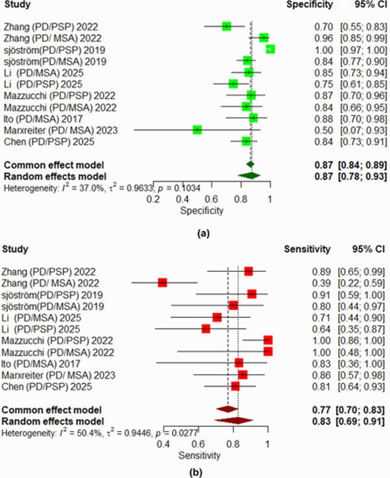
Results: Twelve studies met the inclusion criteria, including 626 patients with PD, 160 patients with MSA and 176 patients with PSP across the 12 studies, along with 256 healthy controls. PD patients consistently showed increased substantia nigra (SN) susceptibility compared to atypical Parkinsonian syndrome (APS), (pooled SMD = 1.24, 95% CI: 0.89-1.90; I² = 52 %). MSA demonstrated significant putaminal susceptibility increases (SMD = 1.00, 95 % CI: 0.49-1.52; I² = 33 %), and AUC of 0.83 (95% CI: 0.74-0.91). PSP exhibited the most pronounced red nucleus (RN) susceptibility elevation (SMD = 1.75, 95 % CI: 0.84-2.67; I² = 92.7%), with AUC of 0.89, (95% CI: 0.81-0.97). Publication bias was not statistically significant in the PD vs. Control or PSP vs. PD comparisons. However, evidence of potential small-study effects was observed in the putaminal MSA analyses (P < 0.05).
Conclusions: QSM demonstrates disorder-specific patterns of iron deposition in Parkinsonian syndromes, with SN in PD, putamen in MSA, and RN in PSP showing the strongest and most consistent signals. However, the use of different pulse sequences, different scanner vendors, and different field strengths may influence susceptibility measurements. Integration of QSM with clinical data and standardization of QSM acquisition and processing protocols, is recommended to improve reliability and enhance its potential value in differentiating parkinsonian syndromes.



ارسال نظر