

Sonographic Correlations With Histological Grade and Biomarker Profiles in Breast Invasive Ductal Carcinoma
Background: Invasive ductal carcinoma (IDC), the most common breast cancer subtype, exhibits significant heterogeneity, limiting traditional prognostic markers. Molecular profiles improve precision, but imaging features may also reflect tumor biology.
Aims: This study evaluates the predictive potential and clinical applicability of ultrasound features for determining tumor grade and molecular profiles in IDC.
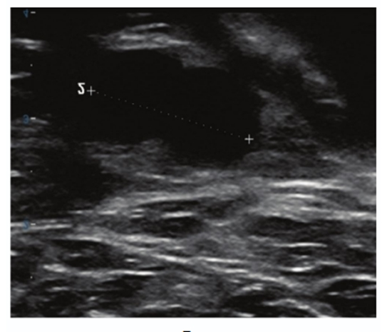
Methods and results: A blinded radiologist retrospectively analyzed 109 IDC cases using the American College of Radiology (ACR) Breast Imaging Reporting and Data System (BI-RADS) classification, evaluating ultrasound features such as lesion shape, margins, orientation, echo pattern, calcifications, vascularity, and lymph node involvement. Tumors were graded histologically (Scarff-Bloom-Richardson system) as low (grades 1 and 2) or high (grade 3). Immunohistochemistry determined estrogen receptor (ER), progesterone receptor (PR), human epidermal growth factor receptor 2 (HER2), and Ki-67 status. ER and PR positivity were defined as > 10% nuclear staining, HER2 graded on a 0-3+ scale, and Ki-67 positivity as ≥ 10% staining. Statistical analyses, including logistic and linear regression, examined correlations between ultrasound features and histological/molecular profiles. Among 109 women (mean age 48.4 ± 12.5 years), the mean tumor length and width were 21.83 ± 11.22 mm and 15.3 ± 6.97 mm, respectively. Histopathological grading revealed that grade 2 tumors were predominant (51%), while grade 1 and grade 3 tumors were observed in 25% and 24% of cases, respectively. ER and PR positivity were observed in 76.4% and 67.6% of cases, respectively. High-grade tumors were significantly associated with ER and PR negativity (p-value < 0.05). Ultrasound features associated with high-grade tumors included larger tumor length (p-value = 0.029). ER positive tumors had smaller axillary lymph nodes (p-value < 0.05). Likewise, PR positive tumors exhibited smaller suspicious axillary lymph nodes compared to PR negative cases (p-value = 0.004).
Conclusion: Sonographic features may correlate with histological grades and hormone receptor statuses in breast IDC, suggesting that ultrasound could aid in predictive assessment.


ارسال نظر