


Differentiation of edematous, tumoral and normal areas of brain using diffusion tensor and neurite orientation dispersion and density imaging
Background
Presurigical planning for glioma tumor resection and radiotherapy treatment require proper delineation of tumoral and peritumoral areas of brain. Diffusion tensor imaging (DTI) is the most common mathematical model applied for diffusion weighted MRI data. Neurite orientation dispersion and density imaging (NODDI) is another mathematical model for DWI data modeling.
Objective
We studied whether extracted parameters of DTI, and NODDI models can be used to differentiate between edematous, tumoral, and normal areas in brain white matter (WM).
Material and Methods
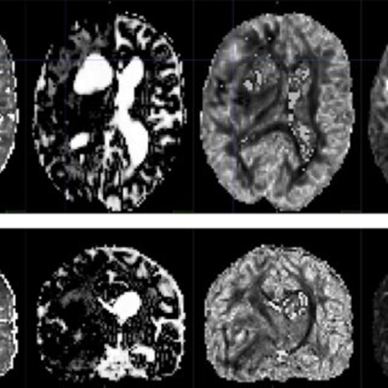
12 patients with peritumoral edema underwent 3T multi-shell diffusion imaging with b-values of 1000 and 2000 smm-2 in 30 and 64 gradient directions, respectively. We fitted DTI and NODDI to data in manually drawn regions of interest and used their derived parameters to characterize edematous, tumoral and normal brain areas.
Results
We found that DTI parameters fractional anisotropy (FA), mean diffusivity (MD), axial diffusivity (AD) and radial diffusivity (RD) all significantly differentiated edematous from contralateral normal brain WM (p<0.005). However, only FA was found to distinguish between edematous WM fibers and tumor invaded fibers (p = 0.001). Among NODDI parameters, the intracellular volume fraction (ficvf) had the best distinguishing power with (p = 0.001) compared with the isotropic volume fraction (fiso), the orientation dispersion index (odi), and the concentration parameter of Watson distribution (κ), while comparing fibers inside normal, tumoral, and edematous areas.
Conclusion
The combination of two diffusion based methods, i.e. DTI and NODDI parameters can distinguish and characterize WM fibers involved in edematus, tumoral, and normal brain areas with reasonable confidence. Further studies will be required to improve the detectability of WM fibers inside the solid tumor if they hypothetically exist in tumoral parenchyma.


ارسال به دوستان