



Ultrasound and shear-wave elastography as a tool for evaluating response to treatment of morphea: a preliminary report of a prospective study
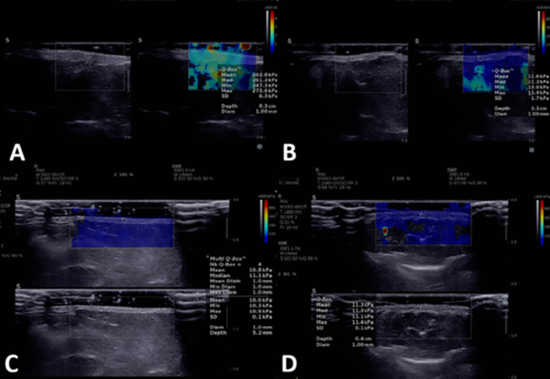
Morphea is an autoimmune connective tissue disorder with no established objective biomarkers for treatment monitoring. Due to the subjectivity of current clinical scoring systems, this study investigates the efficacy of grayscale ultrasound, color Doppler, and shear-wave elastography (SWE) in assessing treatment response and correlating with clinical scores. In this prospective cohort study newly biopsy-confirmed patients with at least one clinically active plaque were followed for 6 months by Color Doppler ultrasound, shear wave elastography, and clinical scoring. The most clinically probable active lesion in each patient was marked and compared to adjacent or contralateral normal skin as control. Eighteen patients with Plaque-type/linear morphea were examined. After six months, dermal thickness as well as mean clinical scores decreased significantly (p < 0.05), while hypodermal thickness decreased and elasticity slightly increased, but it was not significant (p > 0.05). Localized Scleroderma Activity Index (LoSAI) and Localized Scleroderma Damage Index (LoSDI) decreased from 4.28 ± 3.28 to 1.44 ± 1.75 and 11.39 ± 7.38 to 9.56 ± 6.77, respectively. Hypodermal thickness correlated negatively with Physician Global Assessment of Damage (PGA-D) (=-0.410, p = 0.027) and positively with LoSAI ( =0.513, p = 0.004). Additionally, hypodermis thickness was positively correlated with PGA-A before treatment. Furthermore, hypodermal stiffness after treatment was positively correlated with LoSDI. In terms of disease activity assessments, SWE and color Doppler showed poor inter-rater agreement between radiologists and dermatologists (kappa = 0.053). In this preliminary study (N = 18), dermal thickness measured via high-frequency ultrasound may serve as an objective marker for treatment response in morphea. While color Doppler and shear wave elastography (SWE) demonstrated potential as adjunctive tools, their clinical utility remains exploratory and requires validation in larger cohorts.


ارسال نظر