Lower Gray Matter Density Can Be Traceable in Chronic Heroin Dep
https://adir.tums.ac.ir//article/Lower-Gray-Matter-Density-in-the-Anterior-Cingulate-Cortex-and-Putamen-Can-Be-Traceable-in-Chronic-Heroin-Dependents-After-Over-Three-Months-of-Successful-Abstinence
Lower Gray Matter Density in the Anterior Cingulate Cortex and Putamen Can Be Traceable in Chronic Heroin Dependents After Over Three Months of Successful Abstinence Background In recent years, a few studies have addressed the effect of chronic heroin use on brain structure with respect to volume and shape; however, the literature in this field is sparse and further studies are necessary to generate robust replications. Objectives In this study, we intended to assess gray matter density (GMD) di

Computer-aided detection of breast lesions in DCE-MRI
https://adir.tums.ac.ir//article/Computer-aided-detection-of-breast-lesions-in-DCE⁄MRI-using-region-growing-based-on-fuzzy-C-means-clustering-and-vesselness-filter
Computer aided detection of breast lesions in DCE/MRI using region growing based on fuzzy C means clustering and vesselness filter A computer-aided detection (CAD) system is introduced in this paper for detection of breast lesions in dynamic contrast-enhanced magnetic resonance imaging (DCE-MRI). The proposed CAD system firstly compensates motion artifacts and segments the breast region. Then, the potential lesion voxels are detected and used as the initial seed points for the seeded region-grow
Coronary Artery Three-Dimensional Steady-State Free Precession
https://adir.tums.ac.ir//article/Diagnostic-Efficacy-of-Coronary-Artery-Three-Dimensional-Steady-State-Free-Precession-Magnetic-Resonance-Angiography-in-Comparison-with-Invasive-Coronary-Angiography-for-Detecting-Coronary-Artery-Disease
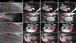
Diagnostic Efficacy of Coronary Artery Three Dimensional Steady State Free Precession Magnetic Resonance Angiography in Comparison with Invasive Coronary Angiography for Detecting Coronary Artery Disease To assess the diagnostic value of three-dimensional steady-state free precession magnetic resonance angiography (3D-SSFP MRA) for detecting coronary artery disease (CAD) Purpose To assess the diagnostic value of three-dimensional steady-state free precession magnetic resonance angiography (3D-SS

1H-MRSI and DWI for Non-invasive Grading of Brain Gliomas
https://adir.tums.ac.ir//Research-Article/Efficacy-of-1H⁄MRSI-and-DWI-for-Non-invasive-Grading-of-Brain-Gliomas
Efficacy of 1H/MRSI and DWI for Non invasive Grading of Brain Gliomas Background Distinguishing low-grade from high-grade gliomas can aid in optimal treatment planning and prognostication. Diffusion-weighted imaging (DWI) and magnetic resonance spectroscopy (MRS) have been applied in several studies for non-invasive glioma grading. However, these studies focused on limited aspects of these imaging techniques and used different study setups, resulting in occasionally inconsistent and incomparable

Gastroesophageal Varices in a Non-Cirrhotic Female with Left Is
https://adir.tums.ac.ir//Research-Article/Extensive-Gastroesophageal-Varices-in-a-Non-Cirrhotic-Female-with-Left-Isomerism:-A-Case-Report-and-Literature-Review
Extensive Gastroesophageal Varices in a Non Cirrhotic Female with Left Isomerism A Case Report and Literature Review Heterotaxy syndrome with polysplenia is a congenital abnormality with malposition of the visceral organs and multiple associated abnormalities. The majority of affected individuals die during childhood due to severe cardiovascular anomalies. However, affected individuals who reach adulthood are usually asymptomatic. Anomalies of the abdominal venous system, including interruption

Cortical Brain Mapping Using fmri
https://adir.tums.ac.ir//news/The-Human-Olfactory-System:-Cortical-Brain-Mapping-Using-fmri
The Human Olfactory System Background Functional magnetic resonance imaging (fMRI) is a non-invasive and convenient method of mapping brain activity associated with the human sensory systems. Among these systems there is a lack of data from olfactory studies, which could be attributed to technical difficulties in odor delivery during scanning. The current study took advantage of an olfactometer to evaluate brain activity during the odor-smelling process. Objectives This study aimed to investigat
Magnetic Resonance Imaging Features of Adenosis in the Breast
https://adir.tums.ac.ir//Research-Article/Magnetic-Resonance-Imaging-Features-of-Adenosis-in-the-Breast-(vol-18,-pg-187,-2015)
(vol 18, pg 187, 2015) In this study, we aim to describe the features of breast adenosis lesions with suspicious or borderline findings on dynamic contrast-enhanced magnetic resonance imaging (DCE-MRI) Purpose Adenosis lesions of the breast, including sclerosing adenosis and adenosis tumors, are a group of benign proliferative disorders that may mimic the features of malignancy on imaging. In this study, we aim to describe the features of breast adenosis lesions with suspicious or borderline fin
Localized-atlas-based segmentation of breast MRI in a decision-m
https://adir.tums.ac.ir//news/Localized-atlas-based-segmentation-of-breast-MRI-in-a-decision-making-framework
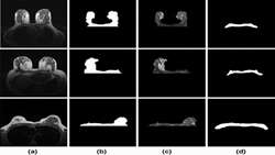
Localized atlas based segmentation of breast MRI in a decision making framework Breast-region segmentation is an important step for density estimation and Computer-Aided Diagnosis (CAD) systems in Magnetic Resonance Imaging (MRI) Breast-region segmentation is an important step for density estimation and Computer-Aided Diagnosis (CAD) systems in Magnetic Resonance Imaging (MRI). Detection of breast-chest wall boundary is often a difficult task due to similarity between gray-level values of fibrog
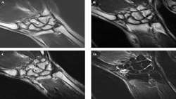
wrist Joint
https://adir.tums.ac.ir//news/Comparison-of-Quantitative-Assessment-of-BLADE-and-Isotropic-Three-Dimensional-Fast-Spin-Echo-Cube-(3D-T2-SPACE)-Sequences-with-Conventional-Protocols-of-wrist-Joint-at-3-Tesla-Magnetic-Resonance-Imaging
Comparison of Quantitative Assessment of BLADE and Isotropic Three Dimensional Fast Spin Echo Cube (3D T2 SPACE) Sequences with Conventional Protocols of at 3 Tesla Magnetic Resonance Imaging Background Magnetic resonance imaging (MRI) of the wrist joint is a useful method in the diagnosis of triangular fibrocartilage complex (TFCC), ligaments and tendons, peripheral nerves, cartilage and carpal tunnel syndrome. However, the evaluation of these small anatomical structures is a topic of investiga
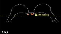
Breast-region segmentation in MRI using chest region atlas and S
https://adir.tums.ac.ir//Research-Article/Breast-region-segmentation-in-MRI-using-chest-region-atlas-and-SVM
Breast region segmentation in MRI using chest region atlas and SVM An important step for computerized analysis of breast magnetic resonance imaging (MRI) is segmentation of the breast region. Due to the similar signal intensity of broglandular tissue and the chest wall, the segmentation process is difficult for breasts with broglandular tissue connected to the chest wall. In order to overcome this challenge, a new framework is presented that relies on a chest region atlas. The proposed method rs