Assessment of pathological response using PET/MRI
https://adir.tums.ac.ir//article/The-role-of-18F⁄FDG-PET⁄MRI-in-assessing-pathological-complete-response-to-neoadjuvant-chemotherapy-in-patients-with-breast-cancer:-a-systematic-review-and-meta-analysis
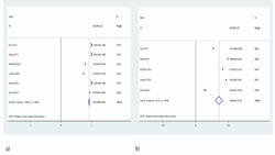
The role of 18F/FDG PET/MRI in assessing pathological complete response to neoadjuvant chemotherapy in patients with breast cancer a systematic review and meta analysis Background and aim The present study aimed to evaluate the use of 18F-2-[18F]-fluoro-2-deoxy-d-glucose (FDG) PET/MRI (Positron emission tomography-computed tomography) in predicting the pathological response to neoadjuvant chemotherapy (NAC) in patients with breast cancer (BC) compared to the use of MRI (Magnetic Resonance Imagin

بررسی تغییرات سیگنال MRI Dentate nucleus در بیماران مبتلا به MS
https://adir.tums.ac.ir//news/بررسی-تغییرات-سیگنال-MRI-Dentate-nucleus-در-بیماران-مبتلا-به-مالتیپل-اسکلروزیس(-MS)-با-دپوزیشن-قابل-توجه-گادولینیوم-در-Dentate-nucleusبا-استفاده-از-سکانس-های-DCE,SWI-در-مقایسه-با-بیماران-ام-اس-فاقد-رسوب-گادولینیوم
بررسی تغییرات سیگنال MRI Dentate nucleus در بیماران مبتلا به مالتیپل اسکلروزیس( MS) با دپوزیشن قابل توجه گادولینیوم در Dentate nucleusبا استفاده از سکانس های DCE,SWI در مقایسه با بیماران ام اس فاقد رسوب گادولینیوم کد خبر 290607 کلمات کلیدی mri_msdentate_nucleus مدیر سیستم 0 * *

یافته های Fetal brain MRI در جنین های IUGR
https://adir.tums.ac.ir//projects/مقایسه-یافته-های-Fetal-brain-MRI-در-جنین-های-IUGR-در-دو-گروه-Clinically-significant-و-Clinically-non-significant
مقایسهدر دو گروه Clinically significant و Clinically non significant کد خبر 290600 کلمات کلیدی fetal_brain_mri_iugr_ مدیر سایت 0 * *
بررسی مقایسه ایی TT-TG distance در MRI
https://adir.tums.ac.ir//news/بررسی-مقایسه-ایی-TT⁄TG-distance-در-MRI-بیماران-مبتلا-به-درد-پتلوفمورال-ایزوله-با-گروه-کنترل
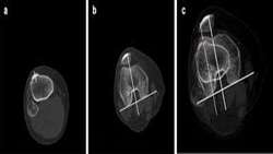
بررسی مقایسه ایی TT/TG distance در MRI بیماران مبتلا به درد پتلوفمورال ایزوله با گروه کنترل کد خبر 290586 کلمات کلیدی mri_tt-tg_distanceپتلوفمورال مدیر سیستم 0 * *

fetal ultrasonography and intra uterine mri brain
https://adir.tums.ac.ir//Research-Article/Fetal-brain-imaging:-A-comparison-between-fetal-ultrasonography-and-intra-uterine-magnetic-resonance-imaging-(a-systematic-review-and-meta-analysis)
Fetal brain imaging A comparison between fetal ultrasonography and intra uterine magnetic resonance imaging (a systematic review and meta analysis) Objective The aim of this study was to compare ultrasound (US) and intra uterine MRI (IUMRI) of the brain in the diagnosis of fetal brain abnormalities. Methods The present systematic review is done based on guidelines for preferred reporting items for systematic reviews and meta-analysis. All major articles comparing fetal US with IUMRI in fetuses w

Ultrasound and mri features of fetal intracranial cystic
https://adir.tums.ac.ir//news/Ultrasound-and-magnetic-resonance-imaging-features-of-fetal-intracranial-cystic-lesions:-A-pictorial-essay
Ultrasound and magnetic resonance imaging features of fetal intracranial cystic lesions A pictorial essay This pictorial essay focuses on ultrasound (US) and magnetic resonance imaging (MRI) features of fetal intracranial cysts. Intracranial cysts are common findings in prenatal imaging, and if great attention is paid to their size, location, and imaging features, they can be diagnosed accurately. They are usually detected by fetal ultrasound exams. However, when ultrasound data on cystic lesion
.png)
Discordance Between Clinical Symptoms and MRI in Pelvic Floor
https://adir.tums.ac.ir//news/The-High-Rate-of-Discordance-Between-Clinical-Symptoms-and-MRI-Findings-in-Patients-with-Pelvic-Floor-Dysfunction/جستجو
The High Rate of Discordance Between Clinical Symptoms and MRI Findings in Patients with Pelvic Floor Dysfunction Abstract Background & Objective Pelvic floor dysfunctions (PFD) are common disorders among women and affect about 50% of them over 50. About 400,000 American women undergo surgery due to the severity of these disorders. The aim of this study was to investigate the correlation of patients' symptoms with both static and dynamic MRI findings.Materials & Methods In this study, we
Contrast Enhanced Abdominopelvic mri on Expression and Methylati
https://adir.tums.ac.ir//article/Y-The-Effect-of-Contrast-Enhanced-Abdominopelvic-Magnetic-Resonance-Imaging-on-Expression-and-Methylation-
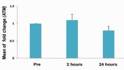
Y The Effect of Contrast Enhanced Abdominopelvic Magnetic Resonance Imaging on Expression and Methylation Objective To evaluate the effect of contrast enhanced abdominopelvic magnetic resonance imaging (MRI), using a 3 Tesla scanner, on expression and methylation level of ATM and AKT genes in human peripheral blood lymphocytes. Materials and Methods In this prospective in vivo study, blood samples were obtained from 20 volunteer patients with mean age of 43 ± 8 years (range 32-68 years) b
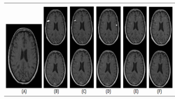
Gadovist and Magnevist in Brain mri of ms
https://adir.tums.ac.ir//news/Comparison-of-Gadovist-and-Magnevist-in-Brain-Magnetic-Resonance-Imaging-of-Multiple-Sclerosis-Patients-with-an-Acute-Attack
Comparison of Gadovist and Magnevist in Brain Magnetic Resonance Imaging of Multiple Sclerosis Patients with an Acute Attack Background The use of an appropriate contrast agent performs a major role in brain Magnetic resonance imaging (MRI) of Multiple sclerosis (MS) patients. Objectives The present study aimed to make a comparison between the diagnostic values of Gadovist and Magnevist considering the successive imaging times in contrast-enhanced brain MRI of MS patients. Methods A total

Multi-parametric MRI for Parotid Tumors
https://adir.tums.ac.ir//news/Radiomic-Analysis-of-Multi-parametric-MR-Images-(MRI)-for-Classification-of-Parotid-Tumors-Running-Title:-Multi-parametric-MRI-for-Parotid-Tumors
Radiomic Analysis of Multi parametric MR Images (MRI) for Classification of Parotid Tumors Running Title Multi parametric MRI for Parotid Tumors Objective To determine the border of glial tumors by diffusion weighted imaging (DWI), apparent diffusion co-efficient (ADC), magnetic resonance spectroscopy (MRS) and perfusion brain MRI. Patients and methods Ten patients with brain gliomas were enrolled [mean age 35.3 ± 13.2, range 20-62]. Conventional MRI was performed for all patients. Beside