Multi-parametric MRI for Parotid Tumors
https://adir.tums.ac.ir//news/Radiomic-Analysis-of-Multi-parametric-MR-Images-(MRI)-for-Classification-of-Parotid-Tumors-Running-Title:-Multi-parametric-MRI-for-Parotid-Tumors
Radiomic Analysis of Multi parametric MR Images (MRI) for Classification of Parotid Tumors Running Title Multi parametric MRI for Parotid Tumors Objective To determine the border of glial tumors by diffusion weighted imaging (DWI), apparent diffusion co-efficient (ADC), magnetic resonance spectroscopy (MRS) and perfusion brain MRI. Patients and methods Ten patients with brain gliomas were enrolled [mean age 35.3 ± 13.2, range 20-62]. Conventional MRI was performed for all patients. Beside
.png)
Diagnostic Algorithm using Multi-parametric MRI Myometrial Tumo
https://adir.tums.ac.ir//article/A-Diagnostic-Algorithm-using-Multi-parametric-MRI-to-Differentiate-Benign-from-Malignant-Myometrial-Tumors:-Machine-Learning-Method
A Diagnostic Algorithm using Multi parametric MRI to Differentiate Benign from Malignant Myometrial Tumors Machine Learning Method This study aimed to develop a diagnostic algorithm for preoperative differentiating uterine sarcoma from leiomyoma through a supervised machine-learning method using multi-parametric MRI. A total of 65 participants with 105 myometrial tumors were included 84 benign and 21 malignant lesions (belonged to 51 and 14 patients, respectively; based on their postoperative ti
MRI Features of Diffuse Cavernous Hemangioma
https://adir.tums.ac.ir//news/Magnetic-Resonance-Imaging-Features-of-Diffuse-Cavernous-Hemangioma-of-the-Uterus-Misdiagnosed-as-a-Low-Grade-Sarcoma:-A-Case-Report
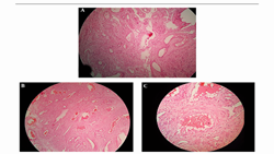
Magnetic Resonance Imaging Features of Diffuse Cavernous Hemangioma of the Uterus Misdiagnosed as a Low Grade Sarcoma A Case Report Uterine cavernous hemangioma is a rare vascular tumor that is more commonly reported as an acquired disease in pregnant women. Rarity of the case impeded the radiologist to be able to find characteristic imaging findings to diagnose the disease before surgery. We report a 40-year-old premenopausal woman with cavernous hemangioma of the uterus that was misdiagnosed a
.jpg)
Detection of Cervical Multiple Sclerosis Lesions
https://adir.tums.ac.ir//news/Comparison-of-Phase-Sensitive-Inversion-Recovery-MRI-with-T2W–TSE-and-STIR-in-the-Detection-of-Cervical-Multiple-Sclerosis-Lesions/266998
Comparison of Phase Sensitive Inversion Recovery MRI with T2W TSE and STIR in the Background Multiple sclerosis (MS) is a chronic inflammatory disease of the central nervous system that causes demyelination or loss of myelin in the white and gray matter of the brain and spinal cord. Objectives In the current study, the phase sensitive inversion recovery (PSIR) sequence was compared with the other sequences (T2-weighted (T2W), short tau inversion recovery (STIR)) to represent the number and con

همبستگی یافته های MRI و CT اسکن در بیماران سیستیک فیبروزیس
https://adir.tums.ac.ir//projects/بررسی-همبستگی-یافته-های-MRI-و-CT-اسکن-در-بیماران-مبتلا-به-سیستیک-فیبروزیس-در-مرکز-طبی-کودکان-شهر-تهران-
بررسی همبستگی یافته های MRI و CT اسکن در بیماران مبتلا به سیستیک فیبروزیس در مرکز طبی کودکان شهر تهران کد خبر 264055 کلمات کلیدی mri_ct_اسکنسیستیک_فیبروزیس مدیر سایت *

الگوها و نتایج non-mass enhancement در MRI پستان
https://adir.tums.ac.ir//news/الگوها--و-نتایج-non-mass-enhancement-در-MRI-پستان-و-تطابق-با-نتایج-پاتولوژی
الگوها و نتایج non mass enhancement در MRI پستان و تطابق با نتایج پاتولوژی کد خبر 263999 کلمات کلیدی mri_پستان مدیر سایت *
.jpg)
یافته های سونوگرافی و MRI و DWI در بدخیمی زمینه ای و ضایعات کبدی
https://adir.tums.ac.ir//projects/بررسی-یافته-های-سونوگرافی-و-MRI-استاندارد-و-DWI-در-بیماران-دارای-بدخیمی-زمینه-ای-و-ضایعات-کبدی-کوچک-تصادفا-کشف-شده-در-سی-تی-اسکن-مراجعه-کننده-به-بیمارستان-امام-خمینی
بررسی یافته های سونوگرافی و MRI استاندارد و DWI در بیماران دارای بدخیمی زمینه ای و ضایعات کبدی کوچک تصادفا کشف شده در سی تی اسکن مراجعه کننده به بیمارستان امام خمینی کد خبر 263838 کلمات کلیدی سونوگرافی_mri_dwi_ضایعات_کبدی مدیر سایت *
.jpg)
ضایعات فوکال کبدی با MRI استاندارد بدون تزریق و دیفیوزیون
https://adir.tums.ac.ir//projects/شناسایی-و-کاراکتریزاسیون-ضایعات-فوکال-کبدی-با-MRI-استاندارد-بدون-تزریق-و-دیفیوزیون-MRI-در-بیماران-مراجعه-کننده-به--مرکز-تصویر-برداری-بیمارستان-امام-خمینی-(ره):-مقایسه-با-دینامیک-MRI
شناسایی و کاراکتریزاسیون MRI در بیماران مراجعه کننده به مرکز تصویر برداری بیمارستان امام خمینی (ره) مقایسه با دینامیک MRI کد خبر 263835 کلمات کلیدی دیفیوزیون_ضایعات_فوکال_کبدیmri_ مدیر سایت *

fmri نواحی حس بویایی در مغز، در سوء مصرف کننده مزمن مت آمفتامین
https://adir.tums.ac.ir//news/ارزیابی-تصویربرداری-عملکردی-نواحی-دخیل-در-حس-بویایی-در-مغز،-در-افراد-سوء-مصرف-کننده-مزمن-مت-آمفتامین
ارزیابی تصویربرداری عملکردی نواحی دخیل در حس بویایی در مغز، در افراد سوء مصرف کننده مزمن مت آمفتامین Activation of the aforementioned brain areas, and the pattern of activation, is largely in accordance with previous published olfactory studies carried out in healthy individuals. کد خبر 263828 کلمات کلیدی fmri_حس_بویاییمت_آمفتامین مدیر سایت *
Diagnostic of MRI and Echocardiography in Constrictive Pericardi
https://adir.tums.ac.ir//article/Diagnostic-Efficacy-of-Magnetic-Resonance-Imaging-and-Echocardiography-in-Diagnosis-of-Constrictive-Pericarditis
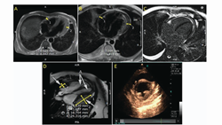
Diagnostic Efficacy of Magnetic Resonance Imaging and Echocardiography in Diagnosis of Constrictive Pericarditis Breast intraductal lesions present a diagnostic challenge due to the diverse spectrum of histologic changes. Vacuum-assisted biopsy (VAB) has evolved as a pivotal diagnostic and therapeutic modality. Yet, concerns about the underestimation of malignancy using VAB persist. This review examines the underestimation rates of Ultrasound-guided VAB (US-VAB) for intraductal lesions and evalu