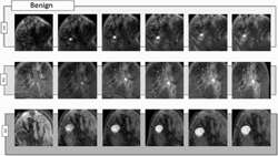
Spatiotemporal features of DCE-MRI for breast cancer diagnosis
https://adir.tums.ac.ir//Research-Article/Spatiotemporal-features-of-DCE-MRI-for-breast-cancer-diagnosis
Spatiotemporal features of DCE MRI for breast cancer diagnosis Background and objective Breast cancer is a major cause of mortality among women if not treated in early stages. Previous works developed non-invasive diagnosis methods using imaging data, focusing on specific sets of features that can be called spatial features or temporal features. However, limited set of features carry limited information, requiring complex classification methods to diagnose the disease. For non-invasive diagnosis
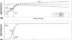
biomarker quantification methodology for DCE-MRI of adnexal mass
https://adir.tums.ac.ir//article/A-one-step-biomarker-quantification-methodology-for-DCE-MRI-of-adnexal-masses:-Capturing-kinetic-pattern-from-early-to-late-enhancement
A one step biomarker quantification methodology for DCE MRI of adnexal masses Capturing kinetic pattern from early to late enhancement Purpose To develop a one-step quantification approach that accounts for joint preprocessing and quantification of whole-range kinetics (early and late-phase washout) of dynamic contrast-enhanced (DCE) MRI of indeterminate adnexal masses. Methods Preoperative DCE-MRI of 43 (24 benign, 19 malignant) sonographically indeterminate adnexal masses were analyzed prospec
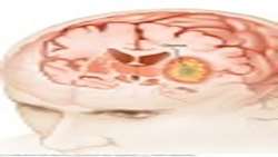
Multimodal MRI in Frameless Navigation Biopsy for Brain Glioma
https://adir.tums.ac.ir//case-report/Multimodal-MRI-Techniques-and-Their-Importance-in-Frameless-Navigation-Biopsy-for-Brain-Glioma
Multimodal MRI Techniques and Their Importance in Frameless Navigation Biopsy for Brain Glioma Gliomas represent 86% of primary intracranial malig- nant tumors. Their most malignant form, glioblastoma multiform (GBM), is either astrocytic, oligodendrocytic, or includes a variety of other histo-pathological subtypes. The best non-invasive imaging modality for neuro- oncologic evaluation of the patients is MRI, which is used for differential diagnosis, biopsy guidance, therapeutic planning, and fo

Computer-aided detection of breast lesions in DCE-MRI
https://adir.tums.ac.ir//article/Computer-aided-detection-of-breast-lesions-in-DCE⁄MRI-using-region-growing-based-on-fuzzy-C-means-clustering-and-vesselness-filter
Computer aided detection of breast lesions in DCE/MRI using region growing based on fuzzy C means clustering and vesselness filter A computer-aided detection (CAD) system is introduced in this paper for detection of breast lesions in dynamic contrast-enhanced magnetic resonance imaging (DCE-MRI). The proposed CAD system firstly compensates motion artifacts and segments the breast region. Then, the potential lesion voxels are detected and used as the initial seed points for the seeded region-grow
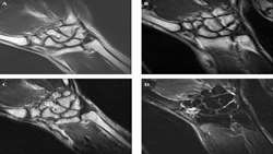
wrist Joint
https://adir.tums.ac.ir//news/Comparison-of-Quantitative-Assessment-of-BLADE-and-Isotropic-Three-Dimensional-Fast-Spin-Echo-Cube-(3D-T2-SPACE)-Sequences-with-Conventional-Protocols-of-wrist-Joint-at-3-Tesla-Magnetic-Resonance-Imaging
Comparison of Quantitative Assessment of BLADE and Isotropic Three Dimensional Fast Spin Echo Cube (3D T2 SPACE) Sequences with Conventional Protocols of at 3 Tesla Magnetic Resonance Imaging Background Magnetic resonance imaging (MRI) of the wrist joint is a useful method in the diagnosis of triangular fibrocartilage complex (TFCC), ligaments and tendons, peripheral nerves, cartilage and carpal tunnel syndrome. However, the evaluation of these small anatomical structures is a topic of investiga

Dynamic Contrast-Enhanced MRI Accurate Classification ol Masses
https://adir.tums.ac.ir//news/f-Complex-AdnexaSemiquantitative-Dynamic-Contrast-Enhanced-MRI-Accurate-Classification-ol-Masses
f Complex AdnexaSemiquantitative Dynamic Contrast Enhanced MRI Accurate Classification ol Masses To identify the best dynamic contrast-enhanced (DCE) magnetic resonance imaging (MRI) descriptive parameters in predicting malignancy of complex ovarian masses, and develop an optimal decision tree for accurate classification of benign and malignant complex ovarian masses Purpose To identify the best dynamic contrast-enhanced (DCE) magnetic resonance imaging (MRI) descriptive parameters in predicting
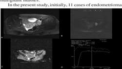
Differentiation of benign from malignant adnexal masses
https://adir.tums.ac.ir//article/Differentiation-of-benign-from-malignant-adnexal-masses-by-functional-3-tesla-MRI-techniques:-diffusion-weighted-imaging-and-time-intensity-curves-of-dynamic-contrast-enhanced-MRI
by functional 3 tesla MRI techniques diffusion weighted imaging and time intensity curves of dynamic contrast enhanced MRI The aim of this study was to evaluate and compare the accuracy of diffusion-weighted imaging (DWI), apparent diffusion coefficient (ADC) value, and time-intensity curve (TIC) type analysis derived from dynamic contrast-enhanced MR imaging (DCE-MRI) in differentiating benign from malignant adnexal masses Background The aim of this study was to evaluate and compare the accurac
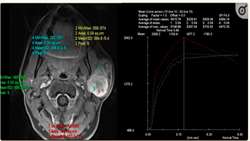
contrast-enhanced MRI for differentiation of major salivary glan
https://adir.tums.ac.ir//news/Dynamic-contrast-enhanced-MRI-for-differentiation-of-major-salivary-glands-neoplasms,-a-3T-MRI-study
Dynamic contrast enhanced MRI for differentiation of major salivary glands neoplasms, a 3T MRI study This study was designed to evaluate the use of dynamic contrast-enhanced MRI (DCE-MRI) for differentiation between malignant, Warthin and benign non-Warthin (BNW) neoplasms of major salivary glands Objectives Pre-operative differentiation of salivary gland neoplasms is of great importance. This study was designed to evaluate the use of dynamic contrast-enhanced MRI (DCE-MRI) for differentiation b

Systematic bone infection detection in axial diabetic foot MRI
https://adir.tums.ac.ir//news/Systematic-bone-infection-detection-in-axial-diabetic-foot-MRI--(Conference-Paper)
(Conference Paper) This paper presents a system for detecting the toe bones in axial diabetic foot MRI and categorizing them Osteomyelitis is an infection of the bone that leads to tissue destruction and often to debility. Early diagnosis of infection is critical, as prompt antibiotic treatment reduces the rate of amputation. Vascular insufficiency and lack of sensation predispose diabetic patients to foot infection that can lead to bone infection. Magnetic Resonance Imaging (MRI) has been shown
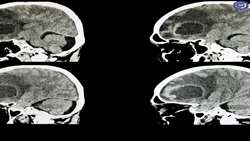
پرفیوژن در درج بندی تومورهای گلیال مغز
https://adir.tums.ac.ir//news/بررسی-حساسیت-و-اختصاصیت--MRI-پرفیوژن-در-درج-بندی-تومورهای-گلیال-مغز-در-بیماران-مراجعه-کننده-به-بیمارستان-امام-خمینی-تهران
بررسی حساسیت و اختصاصیت MRI در بیماران مراجعه کننده به بیمارستان امام خمینی تهران کد خبر 260452 کلمات کلیدی gliomamri_پرفیوژن_ مدیر سایت *