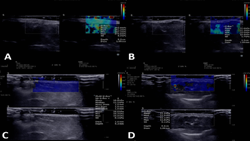
Ultrasound and shear-wave elastography for treatment of morphe
https://adir.tums.ac.ir//Research-Article/Ultrasound-and-shear–wave-elastography-as-a-tool-for-evaluating-response-to-treatment-of-morphea:-a-preliminary-report-of-a-prospective-study
Ultrasound and shear-wave elastography as a tool for evaluating response to treatment of morphea a preliminary report of a prospective study Morphea is an autoimmune connective tissue disorder with no established objective biomarkers for treatment monitoring. Due to the subjectivity of current clinical scoring systems, this study investigates the efficacy of grayscale ultrasound, color Doppler, and shear-wave elastography (SWE) in assessing treatment response and correlating with clinical scores
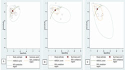
ultrasound attenuation imaging in detecting hepatic steatosis
https://adir.tums.ac.ir//article/Diagnostic-performance-of-ultrasound-attenuation-imaging-in-detecting-hepatic-steatosis
Diagnostic performance of Purpose Hepatic steatosis is a hallmark of metabolic dysfunction-associated steatotic liver disease, and increased hepatic fat (or steatosis) is associated with liver inflammation, fibrosis, and adverse outcomes. While MRI-derived proton density fat fraction (MRI-PDFF) is an accurate noninvasive reference standard, ultrasound attenuation imaging (UAI) offers an accessible cost-effective alternative. We herein systematically review and meta-analyze the accuracy of UAI in

Differences in Ultrasound Characteristics Among Molecular Subtyp
https://adir.tums.ac.ir//news/Differences-in-Ultrasound-Characteristics-Among-Molecular-Subtypes-of-Breast-Cancer:-A-Retrospective-Cross–Sectional-Analysis
es of Breast Cancer A Retrospective Cross-Sectional Analysis Background Breast cancer molecular subtyping is crucial for prognosis and treatment planning, prompting interest in noninvasive ultrasound-based approaches for identifying relevant correlations. Objectives Breast cancer, which is highly prevalent worldwide, comprises several molecular subtypes, including luminal A (LA), luminal B (LB), triple-negative (TN), and human epidermal growth factor receptor 2-positive (HER2+). These subtypes h

Ultrasound findings in the early diagnosis of idiopathic granulo
https://adir.tums.ac.ir//Research-Article/Ultrasound-findings-in-the-early-diagnosis-of-idiopathic-granulomatous-mastitis:-a-large–scale-study
matous mastitis a large-scale study Background Idiopathic granulomatous mastitis (IGM) is a benign but distressing chronic disease. Various etiologies have been proposed for this condition. Although histological examination is used for the definitive diagnosis of the disease, imaging methods including ultrasound (US) are important for achieving correct diagnosis, as well as assisting treatment in some cases. The aim of our study was to identify the most frequent and specific US findings for earl

Ultrasound Radio Frequency Time Series for Tissue Typing
https://adir.tums.ac.ir//Research-Article/Ultrasound-Radio-Frequency-Time-Series-for-Tissue-Typing:-Experiments-on-In–Vivo-Breast-Samples-Using-Texture–Optimized-Features-and-Multi–Origin-Method-of-Classification-(MOMC)
Experiments on In-Vivo Breast Samples Using Texture-Optimized Features and Multi-Origin Method of Classification (MOMC) Objectives One of the most promising auxiliaries for screening breast cancer (BC) is ultrasound (US) radio-frequency (RF) time series. It has the superiority of not requiring any supplementary equipment over other methods. This article sought to propound a machine learning (ML) method for the automated categorization of breast lesions-categorized as benign, probably benign, sus
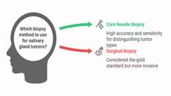
Needle Biopsy in Differentiating Salivary Gland Lesions
https://adir.tums.ac.ir//research-article/The-Value-of-Ultrasound-guided-Core-Needle-Biopsy-in-Differentiating-Benign-from-Malignant-Salivary-Gland-Lesions
The Value of Ultrasound guided Core Needle Biopsy in Differentiating Benign from Malignant Salivary Gland Lesions Purpose In recent years, core needle biopsy (CNB) technique has received much attention, being used as alternative method of tissue sampling for surgical biopsy of salivary gland tumors (SGTs). The present study aimed to evaluate the value of CNB in differentiating benign from malignant SGTs. Materials and methods Patients with suspected benign or malignant SGTs in imaging were enrol

dl for breast lesion classification by ultrasound Images
https://adir.tums.ac.ir//article/A-novel-deep-learning-model-for-breast-lesion-classification-using-ultrasound-Images:-A-multicenter-data-evaluation
A novel deep learning model for breast lesion classification using ultrasound Images A multicenter data evaluation Purpose Breast cancer is one of the major reasons of death due to cancer in women. Early diagnosis is the most critical key for disease screening, control, and reducing mortality. A robust diagnosis relies on the correct classification of breast lesions. While breast biopsy is referred to as the "gold standard" in assessing both the activity and degree of breast cancer, it is an inv

ultrasound & mri Fetal corpus callosum abnormalities
https://adir.tums.ac.ir//news/Fetal-corpus-callosum-abnormalities:-Ultrasound-and-magnetic-resonance-imaging-role/273714
Fetal corpus callosum abnormalities Ultrasound and magnetic resonance imaging role The corpus callosum (CC) is the major interhemispheric commissure and its abnormalities include agenesis, hypoplasia, and hyperplasia. The CC anomalies are typically related to other central nervous system (CNS) or extra-CNS malformations. The antenatal diagnosis of complete CC agenesis is easy after mid-trimester by ultrasound (US) even in the axial plane. The non-visualization of cavum septum pellucidum and colp

Ultrasound and mri features of fetal intracranial cystic
https://adir.tums.ac.ir//news/Ultrasound-and-magnetic-resonance-imaging-features-of-fetal-intracranial-cystic-lesions:-A-pictorial-essay
Ultrasound and magnetic resonance imaging features of fetal intracranial cystic lesions A pictorial essay This pictorial essay focuses on ultrasound (US) and magnetic resonance imaging (MRI) features of fetal intracranial cysts. Intracranial cysts are common findings in prenatal imaging, and if great attention is paid to their size, location, and imaging features, they can be diagnosed accurately. They are usually detected by fetal ultrasound exams. However, when ultrasound data on cystic lesion
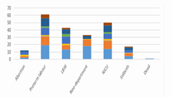
Neonatal Outcomes of Fetuses with Isolated or Multiple Soft Mark
https://adir.tums.ac.ir//article/Neonatal-Outcomes-of-Fetuses-with-Isolated-or-Multiple-Soft-Markers-in-Ultrasound-Screening
ers in Ultrasound Screening Background There is still controversy on the importance of soft markers on the fetus and neonatal outcomes. This study aimed to determine the mentioned outcomes in the fetuses with soft markers detected by ultrasound screening. Methods This prospective study was conducted on 461 pregnant women who were referred to the prenatal clinics of hospitals affiliated with the Tehran University of Medical Sciences, Tehran, Iran, in 2021. The study outcomes included incidence of