Ultrasound and MR Imaging of Fetal Gastrointestinal Abnormalitie
https://adir.tums.ac.ir//Research-Article/A-Spectrum-of-Ultrasound-and-MR-Imaging-of-Fetal-Gastrointestinal-Abnormalities·-Part-2:-Anorectal-Malformation,-Liver,-and-Abdominal-Wall-Anomalies
A Spectrum of s. Part 2 Anorectal Malformation, Liver, and Abdominal Wall Anomalies Background Nasal dorsum augmentation is a highly challenging rhinoplastic procedure. Problems encountered in this surgery, such as residual humps, irregularities, and asymmetry, account for a significant number of revision rhinoplasties. Objectives The aim of this meta-analysis was to assess complication rates and graft harvesting site morbidity and revision rates. Methods We carried out a systematic review of th

ultrasound features and BI-RADS categories of inmalignant breas
https://adir.tums.ac.ir//article/Assessment-of-ultrasound-features-and-BI-RADS-categories-of-inmalignant-breast-masses-Women-≤-40
Assessment of ultrasound features and BI RADS categories of inmalignant breast masses Women ≤ 40 Background To evaluate ultrasound (US) characteristics and BI-RADS (Breast imaging-reporting and data system) of malignant breast masses in women <40 years and to compare with older patients. Methods In a retrospective, descriptive-analytical study, we assessed the US images and BI-RADS category of 78 malignant masses with a final pathology of invasive ductal carcinoma (IDC, NOS type). Results Ove

Ultrasound Measures with POP-Q in Pelvic Organ Prolapse
https://adir.tums.ac.ir//news/Agreement-of-Ultrasound-Measures-with-POP-Q-in-Patients-with-Pelvic-Organ-Prolapse
Agreement of Ultrasound Measures with POP Q in Patients with Pelvic Organ Prolapse Background Ultrasound has emerged as a valuable complimentary tool for assessment of pelvic organ prolapse (POP). Objectives The present study aimed to evaluate the correlation between ultrasound measures and clinical staging in patients with suspected POP. Patients and Methods Forty women with clinical suspicion of POP were enrolled in this cross-sectional study between November 2011 and April 2012. Pelvic organ

reconstruction filters on ultrasound computed tomography image
https://adir.tums.ac.ir//news/Effect-of-reconstruction-filters-on-ultrasound-computed-tomography-image-quality-in-two-different-detector-arrangements:-a-preliminary-simulation-study
Effect of quality in two different detector arrangements a preliminary simulation study Purpose In soft tissues, tumors have generally different sound speeds than normal tissues, so that sound speed images could be used to characterize breast cancer and to study the tumor invasion process. Ultrasound Computed Tomography (UCT) has a great potential to provide quantitative estimations of physical properties of normal and abnormal tissues that provides accurate characterization capability for breas

Intra- and Inter-Rater Reliability of Ultrasound in Plantar Fasc
https://adir.tums.ac.ir//news/Intra-and-Inter–Rater-Reliability-of-Ultrasound-in-Plantar-Fascia-Thickness-Measurement
Intra and Inter-Rater Reliability of Ultrasound in Plantar Fascia Thickness Measurement Background Plantar fascia is a thick connective tissue on the plantar side of the foot, which provides the normal shape of the longitudinal arch. Plantar fasciitis is an inflammation on the medial tuberosity of calcaneus on the inferior side that may cause heel pain. Imaging plays an important role in the diagnosis of heel pain. Ultrasonography has been recognized as an effective imaging tool for assessing pl
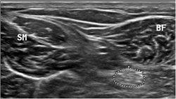
reliability of nerve and muscle morphometric ultrasound
https://adir.tums.ac.ir//article/Test-retest-reliability-of-nerve-and-muscle-morphometric-characteristics-utilizing-ultrasound-imaging-in-individuals-with-unilateral-sciatica-and-controls
Test retest reliability of nerve and muscle morphometric characteristics utilizing ultrasound imaging in individuals with unilateral sciatica and controls Background Ultrasound imaging has been suggested for studying the structure and function of nerves and muscles; however, reliability studies are limited to support the usage. The main aim of this study was to explore the intrarater within-session reliability of evaluating the sciatic nerve and some related muscles morphology by ultrasound imag

bubbles-induced by ultrasound & cholesterol diet for atheroscler
https://adir.tums.ac.ir//Research-Article/Inertial-cavitation-bubbles-induced-by-combined-high-and-low-frequency-ultrasound-accompanied-by-discontinuation-of-cholesterol-rich-diet-for-advanced-atherosclerosis-regression
Inertial cavitation bubbles induced by combined high and low frequency ultrasound accompanied by discontinuation of cholesterol rich diet for advanced atherosclerosis regression Aim Most cases of chest pain and almost all heart attacks are caused by atherosclerosis, a narrowing and hardening of the arteries that results from the accumulation of fatty deposits known as atheromas or plaques. The plaques typically comprise cholesterol- containing low- density lipoproteins (LDLs), smooth muscle cell

Ultrasound in Management of Gestational Trophoblastic Disease
https://adir.tums.ac.ir//Research-Article/Complementary-Role-of-Ultrasound-in-Management-of-Gestational-Trophoblastic-Disease
Complementary Role of To identify an effective method to identify high-risk patients for developing malignancy after molar evacuation Background Transvaginal Ultrasonography is a noninvasive and inexpensive medical imaging tool used for the diagnosis of various diseases. Objectives To identify an effective method to identify high-risk patients for developing malignancy after molar evacuation. Patients and Methods A prospective serial assessment of 19 patients with gestational trophoblastic disea

ultrasound elastography for determining carpal tunnel syndrome
https://adir.tums.ac.ir//news/Application-of-ultrasound-elastography-for-determining-carpal-tunnel-syndrome-severity
Application of severity To evaluate grey-scale and elastography ultrasound imaging findings in patients with CTS compared to nerve conductive studies. Objective To evaluate grey-scale and elastography ultrasound imaging findings in patients with CTS compared to nerve conductive studies. Method Sixty median nerves of 31 cases with confirmed CTS and 44 median nerves in 22 controls (healthy volunteers) who had no clinical evidence of CTS were evaluated. An expert radiologist performed all US evalua

hydrogen peroxide- enhanced endoanal ultrasound in detecting the
https://adir.tums.ac.ir//news/Evaluation-of-hydrogen-peroxide–-enhanced-endoanal-ultrasound-in-detecting-the-internal-orifice-and-type-of-fistula–in–ano
Evaluation of internal orifice and type of fistula-in-ano Hydrogen peroxide-enhanced EUS is a suitable method for detecting the internal orifice of anal fistulas. It can be used for detecting trans-sphincteric fistulas, which are the most common type Background/Objective Accurate delineation of the fistula tract anatomy is necessary for surgical management of anal fistulas. Among different ways to do this, endoanal ultrasound (EUS) is being increasingly used to evaluate patients with anal fistul