

Ultrasonographic evaluation of cosmetic fillers: patterns and frequent complications - A literature review
Purpose: The use of cosmetic fillers has become increasingly popular. Radiologists and sonographers should be aware of the ultrasonographic characteristics of the most commonly used cosmetic fillers and the sonographic features of their frequent complications.
Methods: Ultrasound findings of hyaluronic acid (HA), poly-L-lactic acid (PLLA), calcium hydroxyapatite (CaHA), polymethylmethacrylate (PMMA), polycaprolactone (PCL), silicone oil, polyalkylimide, polyacrylamide (PAAG) and autologous fat were systematically reviewed. Immediate, early, and delayed filler complications-including vascular occlusion, cellulitis and abscess formation, panniculitis, foreign-body granulomatous reaction, fat necrosis, capsular contraction, filler migration, and overfilling-were described with their specific sonographic characteristics.
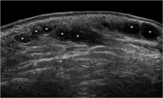
Results: Filler-pattern ultrasound is material-specific: HA-anechoic/hypoechoic, pseudocystic deposits; PLLA-initially hyperechoic, fading over time; PCL-a hypoechoic matrix with hyperechoic comet-tail spots; PMMA-bright echogenic deposits with comet-tail artifacts; CaHA-echoes/shadows depend on dilution/mixing; PAAG-mostly anechoic/hypoechoic and stable over time; silicone-distinctive snowstorm/posterior reverberation; autologous fat-hypoechoic nodules with possible fat necrosis. Vascular occlusion shows absent/reduced Doppler flow; Cellulitis/panniculitis show increased echogenicity, thickened septa, and edema; abscess is an anechoic/hypoechoic lesion with debris and posterior enhancement. Granulomas are hypoechoic nodules with possible calcifications or vascularity. Fat necrosis presents as oil cysts (round anechoic deposits with echogenic borders; calcifications possible). Migration is filler in abnormal locations; overfilling denotes accumulated filler; capsular contracture shows as hyperechoic capsule surrounding deposits.
Conclusion: High-frequency ultrasound, with color Doppler, is the first-line modality for identifying filler type, location, and complications. Routine use improves diagnostic accuracy and patient safety in esthetic medicine and guides therapeutic interventions.


ارسال نظر