Evaluation of plaque detection and optimum time of enhancement in acute attack multiple sclerosis after contrast injection
Magnetic resonance imaging (MRI) is important in the early diagnosis of novel or relapsing multiple sclerosis (MS). In addition, the optimal MRI protocol plays an important role in detecting MS plaques

Background: Magnetic resonance imaging (MRI) is important in the early diagnosis of novel or relapsing multiple sclerosis (MS). In addition, the optimal MRI protocol plays an important role in detecting MS plaques.
Purpose: To find the best time to detect MS plaques on MRI after Gadobutrol injection.
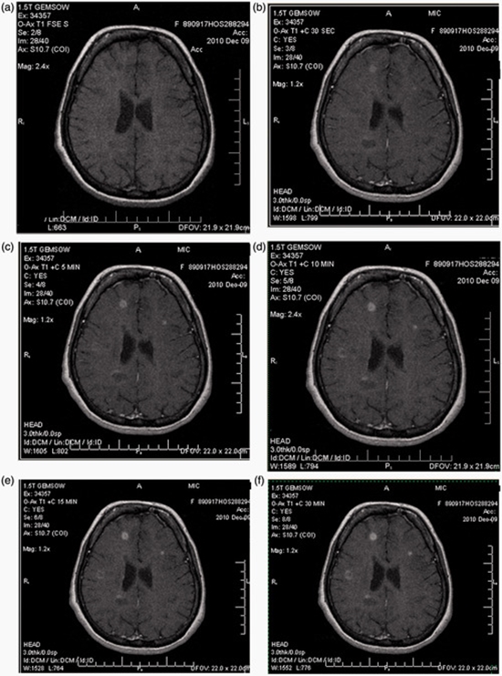
Material and methods: Sixty-two relapsing-remitting type MS patients, (56 women, 6 men) with the mean age of 31 ± 7 years were enrolled into this study. The patients underwent T1-weighted MRI scan without contrast agents. Subsequently, Gadobutrol was injected (0.1 mmol/kg) and MRI scanning was repeated after 30 s, 5, 10, 15, and 30 min of Gadobutrol injection. The size, signal intensity, and enhancement pattern were determined for each plaque by contrast-enhanced T1-weighted images.
Results: Enhancing plaques were seen in 42 out of 62 patients. The mean number of enhancing plaques was 4 ± 8 plaques after 30 s of contrast injection. This figure increased to 7 ± 13 plaques after 15 min and 6 ± 10 plaques after 30 min. The signal intensity and size of plaques increased progressively, and the maximum signal intensity and plaque size were seen after 30 min (P < 0.001).
Conclusion: The maximum number of enhancing plaques in MS patients was detected 15 min after contrast agent administration and the size and signal intensity of the lesions also increased remarkably at this time.


ارسال به دوستان