The Diagnostic Value of CT Angiography in the Diagnosis of Residual Aneurysm After Brain Aneurysm Surgery

Background:
The existence of residual aneurysm after intracranial aneurysm clipping bears the risk of re-bleeding, which worsens with the passage of time. Digital subtraction angiography (DSA) is accepted as the gold standard for evaluation of residual aneurysm, but it is invasive, costly, and serious complications are possible.Objectives:
The aim of this study was to compare DSA to 64-slice CT angiography for assessing residual aneurysm.Patients and Methods:
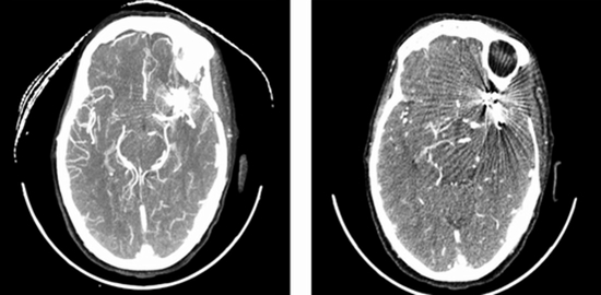
Forty patients with 43 clipped aneurysms from which 36 were torn, were evaluated by DSA after improvement in clinical status, and after a month they were evaluated by 64-slice CT angiography. The pictures were assessed by two neuroradiologists separately, in terms of quality, artifact due to the clips, and the completion of aneurysm closing.Results:
In multislice computed tomographic angiography (MSCT) analysis, 36 pictures (90%) had good quality and four pictures (10%) had poor quality. In case of good quality pictures in MSCT and angiography, the 2- and 3-millimeter residual aneurysms were approved for two patients based on which, sensitivity, feature and positive/negative predictive value for diagnosis of residual aneurysm was 100 for good-quality pictures by MSCT. The level of agreement between the two neuroradiologists was 1 for diagnosing residual aneurysm and 0.86 for vasospasm. The average time for doing MSCT was 12 minutes compared to 45 minutes for DSA angiography, which was cost effective.Conclusion:
CT angiography is a less invasive method with high sensitivity and capabilities for diagnosing residual aneurysm. It is cheaper, quicker and can be accomplished for critical patients. Therefore, it can be taken as the first choice and a replacement for DSA in post-surgery evaluation of patients with clipped brain aneurysm


ارسال به دوستان