

مرکز تحقیقات در یک نگاه
https://adir.tums.ac.ir/adir-overview
مرکز تحقیقات رادیولوژی نوین و تهاجمی مرکز تحقیقات رادیولوژی نوین و تهاجمی (Advanced Diagnostic Interventional Radiology Research Center) در بهار ۱۳۸۸ در مرکز تصویربرداری پزشکی دانشگاه علوم پزشکی و خدمات بهداشتی – درمانی تهران تأسیس شد و موفق به دریافت موافقت اصولی گردید. در اردیبهشت ۱۳۹۲ این مرکز موفق به دریافت موافقت قطعی شد و به عنوان اولین مرکز تحقیقاتی تخصصی در حوزه رادیولوژی در ایران فعالیت خود را ادامه داد. این مرکز با بهرهگیری از تجربه متخصصین رادیولوژی و تجهیزات پیشرفته، بستر
Cell therapy with placenta-derived mesenchymal stem cells
https://adir.tums.ac.ir/news/Cell-therapy-with-placenta-derived-mesenchymal-stem-cells-for-secondary-progressive-multiple-sclerosis-patients-in-a-phase-1-clinical-trial
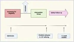
Cell therapy with placenta-derived mesenchymal stem cells Cell therapy with placenta derived mesenchymal stem cells for secondary progressive multiple sclerosis patients in a phase 1 clinical trial --> Mesenchymal stem cell (MSC) has attracted significant attention in clinical research due to their immunomodulatory properties and potential to reduce inflammation in autoimmune disorders, such as multiple sclerosis (MS). This study evaluates the safety and feasibility of placenta-derived MSCs (PLM
Refining MRI protocols for endometriosis
https://adir.tums.ac.ir/news/Refining-MRI-protocols-for-endometriosis:-a-comparative-study-of-abbreviated-and-full-MRI-sequences/293174
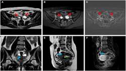
Refining MRI protocols for endometriosis Refining MRI protocols for endometriosis: a comparative study of abbreviated and full MRI sequences --> Objectives: Endometriosis is a significant cause of chronic abdominal pain and infertility in females, often overlooked due to its resemblance to other abdominopelvic pathologies. This study aims to compare the diagnostic performance and agreement rate between an abbreviated MRI protocol (aMRI) and a full MRI protocol (fMRI) for detecting pelvic endomet

Refining MRI protocols for endometriosis
https://adir.tums.ac.ir/news/Refining-MRI-protocols-for-endometriosis:-a-comparative-study-of-abbreviated-and-full-MRI-sequences
Refining MRI protocols for endometriosis Refining MRI protocols for endometriosis: a comparative study of abbreviated and full MRI sequences --> Objectives: Endometriosis is a significant cause of chronic abdominal pain and infertility in females, often overlooked due to its resemblance to other abdominopelvic pathologies. This study aims to compare the diagnostic performance and agreement rate between an abbreviated MRI protocol (aMRI) and a full MRI protocol (fMRI) for detecting pelvic endomet

Resting-State fMRI for Differentiating Cognitive Impairments
https://adir.tums.ac.ir/Research-Article/Quantitative-Assessment-of-Resting-State-Functional-Connectivity-MRI-to-Differentiate-Amnestic-Mild-Cognitive-Impairment,-Late-Onset-Alzheimer’s-Disease-From-Normal-Subjects
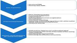
Resting-State fMRI for Differentiating Cognitive Impairments Quantitative Assessment of Resting State Functional Connectivity MRI to Differentiate Amnestic Mild Cognitive Impairment, Late Onset Alzheimer’s Disease From Normal Subjects --> Background: Alzheimer disease (AD) is a neurological disorder with brain network dysfunction. Investigation of the brain network functional connectivity (FC) alterations using resting-state functional MRI (rs-fMRI) can provide valuable information about the bra
Connectivity Evaluation of Resting-State Brain Networks in Alzh
https://adir.tums.ac.ir/Research-Article/Effective-Connectivity-Evaluation-of-Resting-State-Brain-Networks-in-Alzheimer’s-Disease,-Amnestic-Mild-Cognitive-Impairment,-and-Normal-Aging:-An-Exploratory-Study
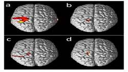
Connectivity Evaluation of Resting-State Brain Networks in Alzh Effective Connectivity Evaluation of Resting State Brain Networks in Alzheimer’s Disease, Amnestic Mild Cognitive Impairment, and Normal Aging: An Exploratory Study --> (1) Background: Alzheimer's disease (AD) is a neurodegenerative disease with a high prevalence. Despite the cognitive tests to diagnose AD, there are pitfalls in early diagnosis. Brain deposition of pathological markers of AD can affect the direction and intensity of
.png)
Preoperative Neuroimaging for Awake Craniotomy
https://adir.tums.ac.ir/article/Preoperative-Conventional-and-Advanced-Neuroimaging-for-Awake-Craniotomy
Preoperative Neuroimaging for Awake Craniotomy Preoperative Conventional and Advanced Neuroimaging for Awake Craniotomy --> Neuroimaging noninvasively provides valuable information about brain structure, physiology, and function. Since the discovery of magnetic resonance imaging (MRI), it has played an efficient role in the diagnosis, treatment planning, and follow-up assessment of brain lesions. Conventional neuroimaging is used to evaluate the lesion location, extension to periphery, size, and
.png)
FMRI to Differentiate Amnestic Mild Cognitive Impairment
https://adir.tums.ac.ir/news/Quantitative-Assessment-of-Resting-State-Functional-Connectivity-MRI-to-Differentiate-Amnestic-Mild-Cognitive-Impairment,-Late-Onset-Alzheimer’s-Disease-From-…
FMRI to Differentiate Amnestic Mild Cognitive Impairment Quantitative Assessment of Resting State Functional Connectivity MRI to Differentiate Amnestic Mild Cognitive Impairment, Late Onset Alzheimer’s Disease From … --> Background: Alzheimer disease (AD) is a neurological disorder with brain network dysfunction. Investigation of the brain network functional connectivity (FC) alterations using resting-state functional MRI (rs-fMRI) can provide valuable information about the brain network pattern
Brain Cortical Activation Wrist Movement
https://adir.tums.ac.ir/news/Brain-Cortical-Activation-during-Imagining-of-the-Wrist-Movement-using-Functional-Near-Infrared-Spectroscopy(fNIRS)
Brain Cortical Activation Wrist Movement Brain Cortical Activation during Imagining of the Wrist Movement using Functional Near Infrared Spectroscopy(fNIRS) --> Background: fNIRS is a useful tool designed to record the changes in the density of blood’s oxygenated hemoglobin (oxyHb) and deoxygenated hemoglobin (deoxyHb) molecules during brain activity. This method has made it possible to evaluate the hemodynamic changes of the brain during neuronal activity in a completely non-aggressive ma
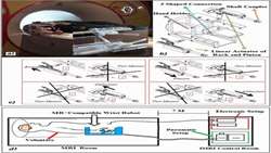
FMRI-Compatible wrist robotic device
https://adir.tums.ac.ir/Research-Article/Novel-FMRI-Compatible-wrist-robotic-device-for-brain-activation-assessment-during-rehabilitation-exercise
FMRI-Compatible wrist robotic device Novel FMRI Compatible wrist robotic device for brain activation assessment during rehabilitation exercise --> Magnetic Resonance Imaging (MRI) can be applied to study the effects of rehabilitation strategies for neuroscience research. An MRI-wrist robot is designed and used as a clinical tool to examine the process of the brain plasticity changes. In this robot, the patient actuation is accomplished with two standard air cylinders, located inside the MRI cham

